. 2024 Nov 29;10(48):eadn6037.
doi: 10.1126/sciadv.adn6037. Epub 2024 Nov 29.

The crystal and cryo-EM structures of PLCγ2 reveal dynamic interdomain recognitions in autoinhibition

Affiliations

The crystal and cryo-EM structures of PLCγ2 reveal dynamic interdomain recognitions in autoinhibition

Young-Cheul Shin et al. Sci Adv. .

Abstract

Phospholipase C gamma 2 (PLCγ2) plays important roles in cell signaling downstream of various membrane receptors. PLCγ2 contains a multidomain inhibitory region critical for its regulation, while it has remained unclear how these domains contribute to PLCγ2 activity modulation. Here we determined three structures of human PLCγ2 in autoinhibited states, which reveal dynamic interactions at the autoinhibition interface, involving the conformational flexibility of the Src homology 3 (SH3) domain in the inhibitory region, and its previously unknown interaction with a carboxyl-terminal helical domain in the core region. We also determined a structure of PLCγ2 bound to the kinase domain of fibroblast growth factor receptor 1 (FGFR1), which demonstrates the recognition of FGFR1 by the nSH2 domain in the inhibitory region of PLCγ2. Our results provide structural insights into PLCγ2 regulation that will facilitate future mechanistic studies to understand the entire activation process.


Figures
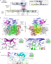
Fig. 1.
Fig. 1.. Crystal structure of human PLCγ2.
(A) Domain architecture of human PLCγ2. Color coding: PH domain, yellow; EF hand, salmon; TIM X/Y, gray; C2 domain, green; spPH domain, magenta; nSH2 domain, light blue; cSH2 domain, slate; SH3 domain, cyan; Y759 linker, bright blue. Newly observed C-terminal helices are noted as H domain and colored in red. Domain boundaries are labeled by amino acid numbers. Construct design of PLCγ2-C is illustrated in the gray box. (B) Crystal structure of human PLCγ2 determined at 2.55-Å resolution (PLCγ2-C), presented in both front (left) and back (right) views. Domains are colored as in (A). Inhibitory region is shown in cartoon representation. The cartoon diagram of the core region is shown embedded in a surface representation. The orange sphere indicates the active site calcium cofactor. (C to E) Interface interactions between the regulatory and core domains of PLCγ2. Domains are colored as in (A). Residues contributing to the interactions are shown in stick representation. Polar interactions are denoted by dashed lines in yellow. Regions of representation are illustrated by the dashed boxes on the complete structure on right, respectively.
Fig. 2.
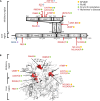
Fig. 2.. Positions and mapping of PLCγ2 mutations and variants.
(A) Positions of gain-of-function point mutations are indicated by red arrows, and the deletions are denoted by red diagonal stripes. Loss-of-function mutation is noted by a dark blue arrow. Disease association is illustrated by filled circles: yellow, APLAID; turquoise blue, PLAID; green, Ibrutinib resistance; purple, Alzheimer’s disease. (B) Mapping of PLCγ2 gain-of-function mutations onto the crystal structure of PLCγ2. Residues at the mutation site are colored in red and shown in sphere representation to label the position of mutation. Label denotes mutations found so far at each position. Disease association is illustrated by filled circles as in (A). Sites for P552R and S1192G are not shown as residues Pro522 and Ser1192 are invisible or absent in the crystal structure, respectively.
Fig. 3.
Fig. 3.. Cryo-EM structure of full-length human PLCγ2 (PLCγ2-F).
(A) Cryo-EM map of PLCγ2-F (3.7-Å resolution) presented in both front (left) and back (right) views. Domains are colored as in Fig. 1A, and the nSH2 domain (in light blue) is visible. (B) Structural model of PLCγ2 with complete inhibitory region. Left, crystal structure of PLCγ2 contains the entire SH3 domain and Y759 linker (highlighted in orange circle) but not the nSH2 domain. Middle, cryo-EM structure of full-length PLCγ2 (PLCγ2-F) has visible nSH2 domain (highlighted in green circle) but misses the vast majority of SH3 and Y759 linker. Right, a structural model of PLCγ2 containing all inhibitory domains was built by overlaying domains visible in both structures. Core region is shown in surface representation in gray. Inhibitory region domains are shown in carton representation and colored as in Fig. 1A.
Fig. 4.
Fig. 4.. Cryo-EM structure of full-length PLCγ2 containing visible C-terminal helical structure, PLCγ2-H.
(A) Cryo-EM structure PLCγ2-H revealed that the C-terminal end forms a double helical structure (newly named H domain) and makes direct contact with SH3 domain in the inhibitory region. Left, cryo-EM map of PLCγ2-H. Domains are colored as in Fig. 1A, and the C-terminal H domain is colored in red. Middle and right, front (middle) and side (right) views of PLCγ2-H structure. C-terminal helices are shown in cartoon representation in red, and the rest of the core region is shown in surface representation in gray. Inhibitory region domains are shown in cartoon representation as colored previously. (B) Conformational flexibility of the SH3 domain in the presence of C-terminal helices. Left, overlay of crystal structure of PLCγ2 (PLCγ2-C) with cryo-EM structure PLCγ2-H highlighted conformational difference of SH3 domain in full-length protein representation. The SH3 domain is shown in cyan in PLCγ2-C, and the SH3 domain and H domain are colored in wheat and red in PLCγ2-H, respectively. The rest of the structures are shown in gray. Middle, focused representation on SH3 domains from the left overlay. Right, overlay of SH3 domain structures from PLCγ2 crystal structure PLCγ2-C (cyan) and cryo-EM structure PLCγ2-H (wheat) by its N-terminal β sheet region (amino acids 767 to 828). The orientation between N- and C-terminal regions of SH3 is illustrated by the relative angel and shown by the dashed arrow. Positions of first (N-) and last (C-) residues of this region are denoted.
Fig. 5.
Fig. 5.. Cryo-EM structure of human PLCγ2 in complex with pFGFR1K.
(A) Cryo-EM map of human PLCγ2/pFGFR1K complex (4.0-Å resolution) presented in both front (left) and back (right) views. PLCγ2 domains are colored as in Fig. 1A, and FGFR1K is colored in orange. (B) Structures of PLCγ2 and PLCγ2/pFGFR1K complex. All shown in cartoon representation and colored as in Fig. 5A. Active site of FGFR1K is noted by a black circle in dashed line. The distance between Y753 in the cSH2-SH3 linker and the active site of FGFR1K is noted by a dashed arrow. PLCγ2 cryo-EM structure only shows PLCγ2-F for illustration.
Fig. 6.
Fig. 6.. Dynamic interactions in autoinhibited states of PLCγ2 and recognition by receptor kinase FGFR1.
The autoinhibited states of PLCγ2 (left) exist in multiple conformations, exhibited by structural flexibility of the SH3 and C-terminal H domains. The recognition sites between cSH2 and C2 domains, and between SH3-spPH linker and TIM barrel, respectively, play indispensable roles. Recognition of PLCγ2 by FGFR1K (middle) is mediated by the nSH2 domain in the inhibitory region, and FGFR1K phosphorylation dependent. This recognition may position PLCγ2 in proximity to the membrane as well as a second FGFR1K nearby, both of which may be involved in the next step of phosphorylation and conformation change of PLCγ2, resulting in the disengagement of the inhibitory region and access of catalytic core to PIP2 substrate membrane (right). Core region is shown in surface representation in gray, in which the TIM barrel that harbors the catalytic center is colored in dark gray. Newly identified H domain is shown in cartoon representation in red. Inhibitory region domains are illustrated in color blocks and colored as in Fig. 1A. FGFR1K associated with PLCγ2 is presented in surface representation in orange, and the rest of the molecule and a second copy of FGFR1 are shown in orange shape blocks. The phosphorylated site of FGFR1K is denoted by “P” in red circle. Plasma membrane bilayer is shown in light gray, and a substrate PIP2 is highlighted in green.

References

    1. Jackson J. T., Mulazzani E., Nutt S. L., Masters S. L., The role of PLCγ2 in immunological disorders, cancer, and neurodegeneration. J. Biol. Chem. 297, 100905 (2021). - PMC - PubMed
    1. Koss H., Bunney T. D., Behjati S., Katan M., Dysfunction of phospholipase Cγ in immune disorders and cancer. Trends Biochem. Sci. 39, 603–611 (2014). - PubMed
    1. Zhou Q., Lee G. S., Brady J., Datta S., Katan M., Sheikh A., Martins M. S., Bunney T. D., Santich B. H., Moir S., Kuhns D. B., Priel D. A. L., Ombrello A., Stone D., Ombrello M. J., Khan J., Milner J. D., Kastner D. L., Aksentijevich I., A hypermorphic missense mutation in PLCG2, encoding phospholipase Cγ2, causes a dominantly inherited autoinflammatory disease with immunodeficiency. Am. J. Hum. Genet. 91, 713–720 (2012). - PMC - PubMed
    1. Neves J. F., Doffinger R., Barcena-Morales G., Martins C., Papapietro O., Plagnol V., Curtis J., Martins M., Kumararatne D., Cordeiro A. I., Neves C., Borrego L. M., Katan M., Nejentsev S., Novel PLCG2 mutation in a patient with APLAID and cutis laxa. Front. Immunol. 9, 2863 (2018). - PMC - PubMed
    1. Martín-Nalda A., Fortuny C., Rey L., Bunney T. D., Alsina L., Esteve-Solé A., Bull D., Anton M. C., Basagaña M., Casals F., Deyá A., García-Prat M., Gimeno R., Juan M., Martinez-Banaclocha H., Martinez-Garcia J. J., Mensa-Vilaró A., Rabionet R., Martin-Begue N., Rudilla F., Yagüe J., Estivill X., García-Patos V., Pujol R. M., Soler-Palacín P., Katan M., Pelegrín P., Colobran R., Vicente A., Arostegui J. I., Severe autoinflammatory manifestations and antibody deficiency due to novel hypermorphic PLCG2 mutations. J. Clin. Immunol. 40, 987–1000 (2020). - PMC - PubMed

Publication types

MeSH terms

Substances

LinkOut - more resources