. 2024 Nov 29;15(1):10388.
doi: 10.1038/s41467-024-54892-w.

Thioredoxin 1 moonlights as a chaperone for an interbacterial ADP-ribosyltransferase toxin

Affiliations

Thioredoxin 1 moonlights as a chaperone for an interbacterial ADP-ribosyltransferase toxin

Baptiste Dumont et al. Nat Commun. .

Abstract

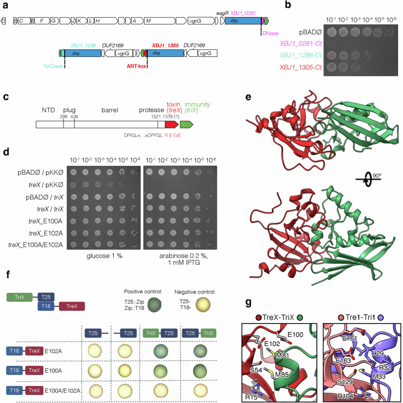
Formation and breakage of disulfide bridges strongly impacts folding and activity of proteins. Thioredoxin 1 (TrxA) is a small, conserved enzyme that reduces disulfide bonds in the bacterial cytosol. In this study, we provide an example of the emergence of a chaperone role for TrxA, which is independent of redox catalysis. We show that the activity of the secreted bacterial ADP-ribosyltransferase (ART) toxin TreX, which does not contain any cysteines, is dependent on TrxA. TreX binds to the reduced form of TrxA via its carboxy-terminal extension to form a soluble and active complex. Structural studies revealed that TreX-like toxins are homologous to Scabin-like ART toxins which possess cysteine residues and form disulfide bridges at the position that superimposes the TrxA binding site in TreX. Our study therefore suggests that thioredoxin 1 evolved alternative functions by maintaining the interaction with cysteine-free substrates.


Conflict of interest statement

Competing interests: The authors declare no competing interests.

Figures

Fig. 1
Fig. 1. The Xenorhadbus bovienii TreX putative ADP-ribosyltransferase toxin is neutralized by the TriX immunity protein.
a Schematic representation of X. bovienii SS-2004 T6SS gene clusters. Rhs are shown in blue, their C-terminal toxic domains (Ct) in purple, cyan, red, and genes encoding their putative cognate immunity proteins are shown in green. The putative activities of the Rhs C-terminal domains are indicated. Genes encoding other T6SS components are indicated as follows: A for TssA, B for TssB, etc. b Rhs C-terminal domains inhibit growth of E. coli when heterologously expressed from pBAD33 vector. Serial dilutions of overnight bacterial cultures carrying vectors coding for different Rhs C-terminal toxins were spotted on LB plates supplemented with 0.2% L-arabinose. c Schematic representation of XBJ1_1305 rhs gene emphasizing the different predicted protein domains—N-terminal domain (NTD) related to protein recruitment to the T6SS, N-terminal plug domain, Rhs fold forming β-roll barrel, aspartyl-transferase (protease) and ADP-ribosyltransferase (ART) toxin domain (red). Pre-toxin sequence carrying DPxGL motifs required for auto-proteolysis leading to toxin release and conserved R-S-ExE residues characteristic to the active site of RSE-clade enzymes are indicated. d Cultures of E. coli MC4100 wild-type cells producing wild-type TreX or its E101A, E103A or E101A/E103A mutants from the pBAD33 vector, and TriX (XBJ1_1304) from the pKK223-3 vector were serially diluted and spotted on LB plates supplemented with 0.2% L-arabinose and 1 mM IPTG. e Crystal structure of TreX (corresponding to XBJ1_1305 C-terminal domain, residues 1380–1518; red) in complex with its immunity protein TriX (green). f Bacterial two hybrid (BACTH) assay. BTH101 reporter cells producing combinations of the indicated T18 and T25 fusion proteins were spotted on LB agar plates supplemented with IPTG and X-Gal. The blue color of the colonies reports an interaction between the two partners. g Zoom-in of the TreX (left) and Tre1 (right) active sites highlighting the position of the inhibiting α-helices of the corresponding immunities (TriX residues 80–85, green; Tri1 residues 27–33, blue, PDB code: 6DRH). The sidechains of the R-S-ExE active site residues of the toxins and the residues of the immunity proteins are indicated.
Fig. 2
Fig. 2. TreX induces cell filamentation.
a Heterologous expression of the TreX toxin induces E. coli filamentation. Microscopy recordings of E. coli cells 60 min after induction of the expression of treX and triX, as indicated. b Schematic representation of the chimeric two component cdiA-cdiB contact dependent growth inhibition system. The treX sequence (residues 1380–1518 of X. boviennii XBJ1_1305, red) is branched to the C-terminus of the EC93 cdiA, downstream the translocation domain. The gene coding for immunity protein TriX (green) is placed downstream to protect the secreting cells. c The chimeric Cdi::TreX-TriX system is functional in bacterial competition against E. coli. Target MG1655 NalR pT5::mCherry cells expressing the triX gene from the pKK223-3-TriX vector are protected (top panel), while those carrying the empty pKK223-3 (middle panel) are killed. Attacker cells expressing the TreX toxin from the pCH vector are shown in cyan, while target cells are shown in cherry, merged with blue for DAPI straining of the nucleoid. Arrows in the zoom-in panel below point to filamenting target cells, resulting from arrested cell division. Scale bars, 5 μm.
Fig. 3
Fig. 3. The reduced form of TrxA is required for TreX activity.
a Mutations of five independent evolved strains resistant to the chimeric CDI::TreX-TriX system selected in bacterial competition. Genes and corresponding mutations are indicated (f.s., frameshift after the residue indicated). Asterisks indicate an incomplete list of mutations identified (see Supplementary Table 4 for the complete list of mutations). b E. coli MC4100 wild-type (WT), trxA, trxA C36A, and trxB mutant strains expressing, or not, treX from the pBAD33 vector, were serially diluted and spotted on LB plates supplemented with 0.2% L-arabinose. c In vitro ADP-ribosylation of FtsZ by the TreX toxin in various conditions. Purified FtsZ was incubated with the TreX toxin, biotinyl-NAD+, wild-type or C36A mutant TrxA, reducing agent DTT or TrxB and NADPH, as indicated, for 15 min at 37 °C. Reactions were then subjected to SDS-PAGE, transfer onto nitrocellulose, and stained with Ponceau Red (top panel). Biotinyl-ADP-ribosylation was detected using streptavidin-alkaline phosphate conjugate (lower panel). Molecular weight markers (in kDa) are indicated on the left.
Fig. 4
Fig. 4. The TreX toxin interacts with TrxA.
a Pulldown assays. Soluble extracts of cells producing the indicated proteins (TreX*ST, TreX inactive toxin (E102A mutant) fused to a Strep tag; TrxAH (His-tagged E. coli TrxA) were subjected to Strep, His, or consecutive Strep and His (Strep + His) affinity purifications. Total (T), soluble (S) and elutions (EL) were separated by SDS-PAGE and stained with Coomassie blue (top panel) or transferred onto nitrocellulose membrane and immunodetected with anti-Strep (middle panel) and anti-His (lower panel) antibodies. The 55-kDa band visible by Coomassie staining was identified as GroEL by mass spectrometry. Molecular weight markers (in kDa) are indicated on the left. b Size-exclusion chromatography analysis of purified thioredoxin TrxA (black line), immunity protein TriX (green line), and TreX*-TriX (blue line), TreX*-TrxA (red line) and TreX*-TriX-TxrA (purple line) complexes. Elution volumes of known molecular weight protein standards are indicated. Peak fractions were subjected to SDS-PAGE and Coomassie blue staining (bottom panel) confirmed the presence of the expected proteins.
Fig. 5
Fig. 5. Structural characterization of the TreX-TriX-TrxA tripartite complex.
a Crystal structure of TreX toxin (red) in complex with its immunity protein TriX (green) and E. coli thioredoxin TrxA (purple). b Zoom-in of the interaction site between the C-terminal residues of the TreX toxin and TrxA, highlighting a series of hydrophobic interactions and hydrogen bonds: TreX T130 forms hydrogen bond with TrxA catalytic C33 (left panel), TreX L132 forms a series of hydrophobic interactions with TrxA W32, and I61 and I76 side chains (middle panel), TreX Y137 sidechain forms hydrogen bond and hydrophobic interactions with TrxA D62 and I61, respectively (right panel). c Pulldown assays. Soluble extracts of cells producing the TreX*ST and TrxAH or their mutated versions were subjected to Strep affinity purification. Total extracts (T) and elutions (EL) were separated by SDS-PAGE and stained with Coomassie blue (top panel) or transferred onto nitrocellulose membrane and immunodetected with anti-Strep (middle panel) and anti-His (lower panel) antibodies. Molecular weight markers (in kDa) are indicated on the left. d In vitro neutralization of the toxin before or after interaction with TrxA. TreX was pre-incubated with the TriX immunity or with TrxA before adding the other components (FtsZ, biotinyl-NAD+, DTT) when indicated with brackets. The immunity protein was also added after the incubation of the toxin and TrxA under reducing conditions, and the reaction was continued for 30 min (right lane). e In vitro ADP-ribosylation of FtsZ with increasing amounts of TreX-TrxA complex reconstituted under denaturing conditions and purified in buffer without DTT. Reactions containing constant amount of FtsZ and biotinyl-NAD+ were mixed with or without the reducing agent DTT for 30 min at 37 °C. The ADP-ribosylation of FtsZ was detected by western-blot using streptavidin-alkaline phosphate conjugate.
Fig. 6
Fig. 6. Conserved interactions of TreX-like toxins with thioredoxins.
a Cladogram of TreX toxin homologs (n = 2195). TreX-like (n = 1027) and Scabin-like (n = 707) clades are suggested based on the absence or the presence of potential disulfide bridge at the C-terminus respectively. b Structure of TreX (red) bound to TrxA (purple) is superimposed to the structure of the toxin Scabin (gray, PDB code: 6VV4). Scabin residues involved in disulfide bridge formation (C176 and C190) align with residues R118 and T130 of TreX, as shown in the zoom-in (right panel). c AlphaFold predicted models of TreX and Scabin-toxin homologs in complex with their cognate thioredoxins. X. bovienii complex (left, colored in red) was solved by X-ray crystallography and aligned to models of homologs from Pseudomonas spp. All AlphaFold prediction statistics are provided in Supplementary Fig. 11 and Supplementary Table 5. d Amino acid sequence alignments of TreX and Scabin toxin homologs. Active site residues are boxed in red, C-terminal extensions interacting with thioredoxins are boxed in blue, cysteines are highlighted in yellow.
Fig. 7
Fig. 7. Proposed model for TrxA binding and toxicity of TreX.
a Structural comparison of the TrxA (purple) binding to its substrate PAPS (left, PDB code: 2O8V), its client T7 polymerase (middle, PDB code: 6P7E), and TreX toxin (right). TrxA is shown in purple, PAPS in pink, T7 polymerase in cyan, DNA in gray, TreX in red, and TriX in green. b Model of secretion of TreX (red), activation by TrxA (purple), and growth inhibition by ADP-ribosylation of FtsZ (blue).

References

    1. Braun, V. & Patzer, S. I. Intercellular communication by related bacterial protein toxins: colicins, contact-dependent inhibitors, and proteins exported by the type VI secretion system. FEMS Microbiol. Lett.345, 13–21 (2013). - PubMed
    1. Cherrak, Y., Flaugnatti, N., Durand, E., Journet, L. & Cascales, E. Structure and activity of the type VI secretion system. Microbiol. Spectr.7, 0031–2019 (2019). - PMC - PubMed
    1. Gallegos-Monterrosa, R. & Coulthurst, S. J. The ecological impact of a bacterial weapon: microbial interactions and the Type VI secretion system. FEMS Microbiol. Rev.45, fuab033 (2021). - PMC - PubMed
    1. Flaugnatti, N. et al. Structural basis for loading and inhibition of a bacterial T6SS phospholipase effector by the VgrG spike. EMBO J.39, e104129 (2020). - PMC - PubMed
    1. Quentin, D. et al. Mechanism of loading and translocation of type VI secretion system effector Tse6. Nat. Microbiol.3, 1142–1152 (2018). - PMC - PubMed

Publication types

MeSH terms

LinkOut - more resources