. 2024 Dec;3(12):1482-1502.
doi: 10.1038/s44161-024-00563-4. Epub 2024 Nov 29.

Immune checkpoint landscape of human atherosclerosis and influence of cardiometabolic factors

Affiliations

Immune checkpoint landscape of human atherosclerosis and influence of cardiometabolic factors

José Gabriel Barcia Durán et al. Nat Cardiovasc Res. 2024 Dec.

Abstract

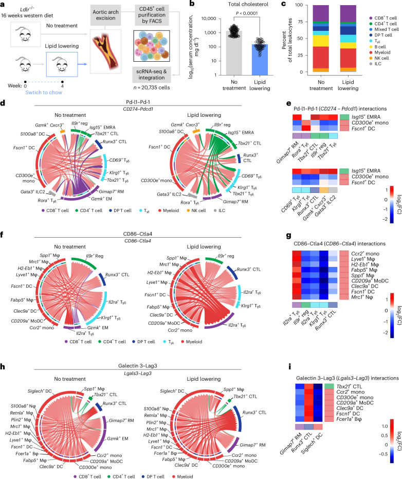
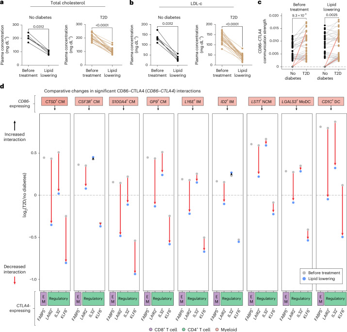
Immune checkpoint inhibitor (ICI) therapies can increase the risk of cardiovascular events in survivors of cancer by worsening atherosclerosis. Here we map the expression of immune checkpoints (ICs) within human carotid and coronary atherosclerotic plaques, revealing a network of immune cell interactions that ICI treatments can unintentionally target in arteries. We identify a population of mature, regulatory CCR7+FSCN1+ dendritic cells, similar to those described in tumors, as a hub of IC-mediated signaling within plaques. Additionally, we show that type 2 diabetes and lipid-lowering therapies alter immune cell interactions through PD-1, CTLA4, LAG3 and other IC targets in clinical development, impacting plaque inflammation. This comprehensive map of the IC interactome in healthy and cardiometabolic disease states provides a framework for understanding the potential adverse and beneficial impacts of approved and investigational ICIs on atherosclerosis, setting the stage for designing ICI strategies that minimize cardiovascular disease risk in cancer survivors.


Conflict of interest statement

Competing interests: The authors declare no competing interests.

Figures

Fig. 1
Fig. 1. Human plaque immune cells display distinct patterns of immune checkpoint gene expression.
a, Schematic depiction of experimental design. scRNA-seq was performed on CD45+ cells from atherosclerotic plaque tissue collected from patients undergoing CEA (n = 22). b, UMAP of scRNA-seq data clustered by cell type. c, Proportions of major immune cell identities per patient. Each dot represents one patient. Boxes represent interquartile ranges; center lines depict medians. Whiskers below and above boxes represent extent of lower and upper quartiles, respectively (n = 22). d, Dot plot of IC gene expression by immune cell subclusters in human atherosclerotic plaques. Dot color represents positive fold change (FC) values in each subcluster compared to the rest (false discovery rate (FDR) < 0.05). Among ‘FDA-approved targets’ are genes encoding direct targets of drugs approved by the US FDA to treat cancer-related indications as well as genes encoding their interaction partners. Among ‘Investigational targets’ are genes encoding ICs currently under investigation as potential cancer immunotherapy targets (ClinicalTrials.gov; 6 December 2023). ICs that do not fit either of these categories appear under ‘Other immune checkpoints’. Mye, myeloid cell; CTL, cytotoxic T lymphocyte; Reg, regulatory; Mono, monocyte; Mφ, macrophage; DC, dendritic cell. Source data
Fig. 2
Fig. 2. Distribution of top immune checkpoint interactions targeted by current immunotherapies among cell populations in the human atherosclerotic plaque.
a,c,e, Chord plots illustrating the strongest interactions (P < 0.05) between: PD-L1 (encoded by CD274) and PD-1 (encoded by PDCD1) (a), CD86 and CTLA4 (c) and galectin 3 (encoded by LGALS3) and LAG3 (e). Direction of ligand → receptor interaction is denoted by chord arrows. b,d,f, Representative immunofluorescent staining of human carotid plaque specimens (left; n = 6–8) and corresponding cell segmentation of phenotypes of interest (top right) for spatial aggregation analysis by NES computation (bottom right). Scale bars, 10 μm. NES > 0 are depicted in red and <0 in blue. Source data
Fig. 3
Fig. 3. CCR7+FSCN1+ dendritic cells are a hub of immune checkpoint communication in human atherosclerotic plaques.
a, Heatmap showing the number of significant (P < 0.05) IC interactions in which ligand-expressing myeloid cells were predicted to engage with T cells within atherosclerotic plaques. P values were calculated by the CellChat package (v.1.1.3) in R (v.4.0.3). b, Violin plots showing normalized transcript abundance and distribution in myeloid cell subclusters engaging in IC interactions with T cells. NE, normalized expression. c, Representative immunofluorescent staining of human carotid plaque specimens (top; n = 3) and proportion of CD11c+ or CD11c+FSCN1+ cells as a percentage of total cells quantified per specimen (bottom). Scale bars, 10 μm. Each dot in the bar plot represents the average of three regions of interest. d, Canonical pathway analysis of differentially expressed transcripts in CCR7+FSCN1+ dendritic cells compared to other myeloid cell populations in human atherosclerotic tissue. Significantly upregulated or downregulated gene pathways terms appear as red or blue dots, respectively. Gray dots indicate pathways below cutoffs for significance (P value < 0.05 and |z-score| > 2), denoted by dotted lines. P values were calculated by the Ingenuity Pathway Analysis software. Some pathway names were edited for brevity. e, Chord plot illustrating the strongest significant IC interactions between ligand-expressing CCR7+FSCN1+ DCs (left) and receptor-expressing subclusters (right). Source data
Fig. 4
Fig. 4. Effect of lipid lowering on the immune checkpoint landscape of atherosclerosis.
a, Schematic depiction of experimental design. scRNA-seq was performed on CD45+ cells from the aortic arches of Ldlr−/− mice subjected to a western diet for 16 weeks with or without an additional 4 weeks of chow diet feeding as a lipid-lowering treatment (n = 10 per group). Raw counts were integrated with equivalent (control or lipid-lowered) specimens from Sharma et al., Afonso et al. and Schlegel et al. for analyses. b, Total cholesterol levels of atherosclerotic mice before and after lipid lowering. P values were determined by unpaired, two-tailed Student’s t-test. c, Proportion of major cell identities in mouse atherosclerotic plaques by scRNA-seq, stratified by treatment. d,f,h, Chord plots illustrating the strongest (P < 0.05) interactions between PD-L1 (encoded by Cd274) and PD-1 (encoded by Pdcd1) (d), CD86 and CTLA4 (f) and galectin 3 (encoded by Lgals3) and LAG3 (h) in mice that received no (left) or lipid-lowering treatment (right). The direction of the ligand → receptor interaction is denoted by chord arrows. Darker chords indicate selected interactions that change with lipid lowering. e,g,i, Heatmaps showing differential PD-L1–PD-1 (e), CD86–CTLA4 (g) and galectin 3–LAG3 (i) interaction probabilities predicted in untreated versus lipid-lowered mice (P < 0.05). P values were calculated by the CellChat package (v.1.1.3) in R (v.4.0.3). Nφ, neutrophil; Bφ, basophil; MoDC, monocyte/dendritic cell. CD8+ T cells are depicted in purple; CD4+ T cells are depicted in green; DP T cells are depicted in dark blue; Tγδ cells are depicted in cyan; myeloid cells are depicted in red; NK cells are depicted in orange; and ILCs are depicted in gray. Source data
Fig. 5
Fig. 5. Immune checkpoint inhibition elicits emergence of FSCN1+ dendritic cells and pro-atherogenic changes in T cells.
a, Schematic depiction of experimental design. Cellular indexing of transcriptomes and epitopes followed by sequencing (CITE-seq) was performed on human PBMCs subjected to anti-CTLA4 or anti-PD-1 treatment for 24 h. b, Proportions of monocyte and dendritic cell subclusters from total myeloid cell compartment, with proportional changes in FSCN1+ dendritic cell abundance highlighted. c, Violin plots illustrating normalized transcript abundance and distribution of indicated genes in myeloid cell subclusters. d, Canonical pathway analysis of differentially expressed transcripts in FSCN1+ dendritic cells compared to other myeloid cell populations. e, Proportions of T cell subclusters from total CD8+ T cell compartment with proportional changes in NKG7+ EM and TFRC+ terminally differentiated EMRA CD8+ T cell abundances highlighted. f, Violin plots illustrating normalized transcript abundance and distribution of indicated genes in CD8+ T cell subclusters. g,h, Canonical pathway analysis of differentially expressed transcripts in NKG7+ EM CD8+ T cells (g) or TFRC+ EMRA CD8+ T cells (h) compared to other CD8+ T cell populations. i, Proportions of T cell subclusters from total CD4+ T cell compartment, with proportional changes in CCR4+ Reg and ENTPD1+ EM CD4+ T cell abundances highlighted. j, Violin plots illustrating normalized transcript abundance and distribution of indicated genes in CD4+ T cell subclusters. k,l, Canonical pathway analysis of differentially expressed transcripts in CCR4+ Reg CD4+ T cells (k) or ENTPD1+ EM CD4+ T cells (l) compared to other CD4+ T cell populations. P values < 0.05 and |z-score| > 2 (d,g,h,k,l). P values were calculated by the Ingenuity Pathway Analysis software. Some Gene Ontology terms were edited for brevity. NE, normalized expression; Tag, aging T cell; TFH, follicular helper T cell; ISG, interferon-stimulated gene; RAR, retinoic acid receptor. m, Schematic depiction of ex vivo anti-PD-1 experiment. CyTOF was performed on human carotid vascular explants subjected to α-PD-1 treatment ex vivo (n = 3). n, Heatmap depicting significant (P < 0.05) T cell marker protein expression by condition. z-score shows scaled average expression. o, Box plot depicting normalized expression of CD45RA in CD8+ or CD4+ T cells derived from control or anti-PD-1-treated explants. Boxes represent interquartile ranges; center lines depict medians. Whiskers below and above boxes represent extent of lower and upper quartiles, respectively. Each dot represents a cell. P values in n and o were determined by two-tailed Wilcoxon rank-sum test (n = 3 per group). Source data
Fig. 6
Fig. 6. PD-1 and CTLA4 interactions are decreased in plaques of type 2 diabetic (T2D) patients.
a, Schematic depiction of IC interaction analyses in atherosclerotic plaque immune cells from T2D versus nondiabetic patients (n = 22). b, Proportion of major cell identities found in human plaques by scRNA-seq, stratified by diabetes status. *P = 0.036. c, Proportions of CD274- or CD86-expressing myeloid cell subclusters engaging in the strongest significant interactions with PD-1 or CTLA4. Boxes represent interquartile ranges; center lines depict medians. Each dot represents a patient. Whiskers below and above boxes represent extent of lower and upper quartiles, respectively. P values in b and c were determined by unpaired, two-tailed Student’s t-tests (n = 9 nondiabetic and n = 13 T2D). d,e, Dot plot of differential PD-L1 (encoded by CD274)–PD-1 (encoded by PDCD1) (c) or CD86–CTLA4 (d) interaction probabilities in T2D versus nondiabetic patients (P < 0.05). P values were calculated by the CellChat package (v.1.1.3) in R (v.4.0.3). Source data
Fig. 7
Fig. 7. Immune-checkpoint interactions in the peripheral blood of patients with type 2 diabetes.
a, Schematic depiction of scRNA-seq analyses of PBMCs from T2D (n = 8) or nondiabetic (n = 4) samples. b, Proportion of major cell identities found in human PBMCs by scRNA-seq. c, PCA of IC gene counts from PBMCs of T2D versus nondiabetic patients. d, Bar plot depicting the relative contribution of co-inhibitory IC interactions to overall cell–cell communication. e, Communication strengths of IC interactions targeted by cancer immunotherapies, ranked in order of probability, with the top 30 interactions depicted as rings. Colors indicate the receptor family, PD-1 (blue), CTLA4 (orange) or LAG3 (green). fh, Chord plots illustrating the strongest (P < 0.05) predicted interactions between CD86 and CTLA4 (f), galectin 3 (encoded by LGALS3) and LAG (g) and αSyn (encoded by SNCA) and LAG3 (h). The direction of the ligand → receptor interaction is denoted by chord arrows. P values were calculated by the CellChat package (v.1.1.3) in R (v.4.0.3). Source data
Fig. 8
Fig. 8. Lipid-lowering treatment is predicted to reduce susceptibility of circulating immune cells to anti-CTLA4 immunotherapy.
a,b, Levels of total cholesterol (a) or LDL-c (b) in individuals with and without T2D, before and after lipid-lowering treatment (n = 12). c, Communication strengths of the strongest (P < 0.05) CD86–CTLA4 interactions before and after lipid-lowering treatment (gray lines; n = 44 interactions). Red lines depict interactions involving CD86-expressing FSCN1+ dendritic cells. Rings represent lack of interaction, hence interaction strength = 0. P values in ac were determined by two-tailed Wilcoxon signed-rank tests. P values for interactions were calculated by the CellChat package (v.1.1.3) in R (v.4.0.3). d, Differential communication strengths of strongest (P < 0.05) CD86–CTLA4 interactions in CD86-expressing myeloid cell subclusters before (gray) and after (light blue) lipid-lowering treatment. Dots represent the mean FC between T2D and nondiabetic patients of the indicated interaction. Dotted line indicates log2(FC) = 0, which is the threshold at which interactions between T2D and nondiabetic patients were predicted to remain unchanged. P values were calculated by the CellChat package (v.1.1.3) in R (v.4.0.3). CM, classical monocyte; IM, intermediate monocyte; NCM, nonclassical monocyte. Source data
Extended Data Fig. 1
Extended Data Fig. 1. Proportions and corresponding gene expression patterns of human atherosclerotic plaque immune cell subclusters by single-cell RNA sequencing.
a–g, Proportions of CD8+ T cell (a), CD4+ T cell (b), Mixed (CD8+ or CD4+) and γδ T (Tγδ) cell (c), double-positive (DP; CD8+ CD4+) T cell (d), B cell (e), myeloid (f), and natural killer (NK) cell and innate lymphoid cell (ILC) (g) subclusters (left) and dot plots showing scaled average expression of corresponding markers (right). EM: effector memory; EMRA: terminally differentiated effector memory re-expressing CD45RA; CM: central memory; RM: resident memory; CTL: cytotoxic T lymphocyte; Reg: regulatory; MAIT: mucosal-associated invariant; NKT: natural killer T cell; Mono: monocyte; Mφ: macrophage; DC: dendritic cell. NE: normalized expression. Source data
Extended Data Fig. 2
Extended Data Fig. 2. Immune checkpoint (IC) gene expression pattern in human coronary artery.
a, Uniform Manifold Approximation and Projection (UMAP) of scRNA-seq data from human coronary arteries (left; n = 6 specimens from 5 subjects) and proportion of each cell type depicted (right), stratified by atherosclerotic disease status: adaptive intimal thickening (AIT, n = 3) or plaque (n = 3). b, Proportions of major immune cell identities sequenced per patient. Each dot represents one specimen. Boxes represent interquartile ranges; center lines depict medians. Whiskers below and above boxes represent extent of lower and upper quartiles, respectively (n = 3 per group). c, Heatmap depicting immune cell marker gene expression by cluster. z-score shows scaled average expression. d, Heatmap of scRNA-seq IC gene expression in AIT (pink), plaque (yellow), both (gray), or neither (white) specimen from human coronary arteries, shown by cell clusters and subclusters. For significantly differentially expressed genes in plaque vs. AIT (FDR < 0.05), fold change (FC) is shown instead. DP: double-positive (CD8+ CD4+); Tγδ: γδ T cell; Mye/M: myeloid cell; NK: natural killer; ILC: innate lymphoid cell; HSPC: hematopoietic stem and progenitor cell; Fibro: fibroblast; MyoF: myofibroblast; SMC: smooth muscle cell; EM: effector memory; EMRA: terminally differentiated effector memory re-expressing CD45RA; CM: central memory; RM: resident memory; CTL: cytotoxic T lymphocyte; Reg: regulatory; MAIT: mucosal-associated invariant; Mono: monocyte; Mφ: macrophage; DC: dendritic cell; FDA: [United States] Food and Drug Administration. Source data
Extended Data Fig. 3
Extended Data Fig. 3. Distribution of significant immune checkpoint (IC) interactions across immune cell clusters and subclusters found in human atherosclerotic plaques by single-cell RNA sequencing.
Heatmap showing the number of significant (p < 0.05) IC interactions predicted between ligand-expressing (y-axis) and receptor-expressing (x-axis) immune cell clusters and subclusters within atherosclerotic plaques. p values were calculated by the CellChat package (v1.1.3) in R (v4.0.3). DP: double-positive (CD8+ CD4+); Tγδ: γδ T cell; NK: natural killer; ILC: innate lymphoid cell; EM: effector memory; EMRA: terminally differentiated effector memory re-expressing CD45RA; CM: central memory; RM: resident memory; CTL: cytotoxic T lymphocyte; Reg: regulatory; MAIT: mucosal-associated invariant; NKT: natural killer T cell; Mono: monocyte; Mφ: macrophage; DC: dendritic cell. Source data
Extended Data Fig. 4
Extended Data Fig. 4. Strength and gene expression profile of clinically relevant immune checkpoint (IC) interactions.
a, Bar plot depicting the relative contribution of predicted IC interactions to overall cell-cell communication among human plaque immune cells. (*) indicates interactions targeted by immunotherapies approved by the U.S. Food and Drug Administration; (†) indicates interactions reported to increase risk of atherosclerotic cardiovascular events,,. b,d,f, Chord plots illustrating the strongest interactions (p < 0.05) between: (b), PD-L2 (encoded by PDCD1LG2) and PD-1 (encoded by PDCD1); (d), CD80 and CTLA4; and (f), α-synuclein (αSyn, encoded by SNCA) and LAG3. Direction of ligand → receptor interaction is denoted by chord arrows. p values were calculated by the CellChat package (v1.1.3) in R (v4.0.3). c,e,g, Communication strengths of: (c), PD-L1– or PD-L2–PD-1 interactions; (e), CD86– or CD80–CTLA4 interactions; and (g), Galectin 3– or αSyn–LAG3 interactions, ranked in order of probability, with the top 30–40 interactions depicted as rings. p values were determined by two-tailed Kolmogorov–Smirnov tests. h, Violin plots illustrating normalized transcript abundance and distribution of IC ligands in myeloid cell subclusters engaging in significant IC interactions. NE: normalized expression. DP: double-positive (CD8+ CD4+); Tγδ: γδ T cell; NK: natural killer; EM: effector memory; EMRA: terminally differentiated effector memory re-expressing CD45RA; RM: resident memory; CTL: cytotoxic T lymphocyte; Reg: regulatory; MAIT: mucosal-associated invariant; NKT: natural killer T cell; Mono: monocyte; Mφ: macrophage; DC: dendritic cell. Source data
Extended Data Fig. 5
Extended Data Fig. 5. Co-inhibitory immune checkpoint interactions targeted by experimental ICIs.
Chord plots illustrating the strongest interactions (p < 0.05) between: (a), HLA-E and and NKG2A (encoded by KLRC1); (b), HMGB1 and TIM3 (encoded by HAVCR2); (c), Galectin 9 (encoded by LGALS9) and TIM3; (d), CD47 and SIRPα (encoded by SIRPA); (e), BTLA and HVEM (encoded by TNFRSF14); and (g), NECTIN2 and TIGIT. Direction of ligand → receptor interaction is denoted by chord arrows. p values were calculated by the CellChat package (v1.1.3) in R (v4.0.3). DP: double-positive (CD8+ CD4+); NK: natural killer; ILC: innate lymphoid cell; EM: effector memory; EMRA: terminally differentiated effector memory re-expressing CD45RA; CM: central memory; RM: resident memory; CTL: cytotoxic T lymphocyte; Reg: regulatory; MAIT: mucosal-associated invariant; NKT: natural killer T cell; Tγδ: γδ T cell; Mono: monocyte; Mφ: macrophage; DC: dendritic cell. CD8+ T cells are depicted in purple; CD4+ T cells are depicted in green; Mixed (CD8+ or CD4+) T cells are depicted in light blue; DP T cells are depicted in dark blue; γδ T (Tγδ) cells are depicted in cyan; B cells are depicted in yellow; myeloid cells are depicted in red; NK cells are depicted in orange; ILC are depicted in gray. f, Communication strengths of NECTIN2, NECTIN3, PVR, or NECTIN4–TIGIT interactions, ranked in order of probability, with the top 30 interactions depicted as rings. p values were determined by two-tailed Kolmogorov–Smirnov tests. Range from lowest to highest p values computed is shown. Source data
Extended Data Fig. 6
Extended Data Fig. 6. Immune checkpoint (IC) interactions in murine atherosclerotic plaque immune cells.
a, Dot plot of immune checkpoint (IC) gene expression by immune cell clusters and subclusters in murine atherosclerotic plaques. Dot color represents fold change (FC) values in each subcluster compared to the rest (FDR < 0.05). b, Bar plot depicting the relative contribution of predicted IC interactions to overall cell-cell communication among murine plaque immune cells. (*) indicates interactions targeted by immunotherapies approved by the U.S. Food and Drug Administration; (†) indicates interactions known to increase risk of cardiovascular events like myocardial infarction and stroke in humans,,. c, Communication strengths of IC interactions targeted by cancer immunotherapies, ranked in order of probability and stratified by treatment, with the top 30 interactions depicted as rings. DP: double-positive (CD8+ CD4+); M: Mixed (CD8+ or CD4+) T cell; Tγδ: γδ T cell; NK: natural killer; ILC: innate lymphoid cell; EM: effector memory; RM: resident memory; Tc17: T cell 17; EMRA: terminally differentiated effector memory re-expressing CD45RA; Th17: T helper 17; Mono: monocyte; Mφ: macrophage; Nφ: neutrophil; Bφ: basophil; MoDC: monocyte/dendritic cell; DC: dendritic cell. Source data
Extended Data Fig. 7
Extended Data Fig. 7. Characterization of anti-inflammatory program in murine atherosclerotic plaque immune cells that results from lipid-lowering treatment.
a–c, Canonical pathway analyses of differentially expressed transcripts in, respectively, myeloid, CD8+ T cells, or CD4+ T cells in lipid-lowered vs. untreated (control) mice. p value < 0.05 and |z-score| > 2. Some GO terms were edited for brevity. d–f, Heatmaps showing normalized transcript abundances of genes contributing to the respective GO terms for, respectively, myeloid, CD8+ T cells, or CD4+ T cells in lipid-lowered (light blue) vs. untreated (control; gray) mice. z-score shows scaled average expression. Source data
Extended Data Fig. 8
Extended Data Fig. 8. Proteomic characterization by cytometry by time-of-flight (CyTOF) of human vascular explants subjected to PD-1 inhibition ex vivo.
af, Box plots depicting normalized expression of indicated markers in CD8+ or CD4+ T cells derived from control or anti (α)-PD-1-treated human carotid vascular explants. Boxes represent interquartile ranges; center lines depict medians. Whiskers below and above boxes represent extent of lower and upper quartiles, respectively. Each dot represents a cell. p values were determined by two-tailed Wilcoxon rank-sum tests (n = 3 per group). Source data
Extended Data Fig. 9
Extended Data Fig. 9. Immune checkpoint (IC) gene expression and interactions in human peripheral blood mononuclear cells (PBMCs).
a, Dot plot of differential IC gene expression by immune cell cluster and subcluster found in human PBMCs from type 2 diabetic (T2D; n = 8) vs. non-diabetic (n = 4) patients. Dot color represents fold change (FC) values in each subcluster compared to the rest (FDR < 0.05). b,c, Chord plots illustrating the strongest (p < 0.05) interactions between: (c) Galectin 9 (encoded by LGALS9) and TIM3 (encoded by HAVCR2) and (d) BTLA and HVEM (encoded by TNFRSF14). Direction of ligand → receptor interaction is denoted by chord arrows. p values were calculated by the CellChat package (v1.1.3) in R (v4.0.3). Unconv: unconventional; NK: natural killer [cell]; ILC: innate lymphoid cell; HSPC: hematopoietic stem and progenitor cell; EM: effector memory; EMRA: terminally differentiated effector memory re-expressing CD45RA; CTL: cytotoxic T lymphocyte; CM: central memory; Reg: regulatory; ExReg: ex-regulatory; NKT: natural killer cell; MAIT: mucosal-associated invariant; Tγδ: γδ T cell; CMono: classical monocyte; IM: intermediate monocyte; NCM: non-classical monocyte; MoDC: monocyte/dendritic cell; DC: dendritic cell. Source data
Extended Data Fig. 10
Extended Data Fig. 10. Comparative changes in significant Galectin 3–LAG3 interactions.
Differential communication strengths of strongest (p < 0.05) Galectin 3 (encoded by LGALS3)–LAG3 interactions in LAG3-expressing myeloid cell subclusters before (gray) and after (light blue) lipid-lowering treatment. Dots represent the mean fold change (FC) between T2D and non-diabetic patients of the indicated interaction. Dotted line indicates log2(FC) = 0, which is the threshold at which interactions between T2D and non-diabetic patients were predicted to remain unchanged. p values were calculated by the CellChat package (v1.1.3) in R (v4.0.3). Tγδ: γδ T cell; EM: effector memory; EMRA: terminally differentiated effector memory re-expressing CD45RA; CTL: cytotoxic T lymphocyte; Tγδ: γδ T cell; CM: classical monocyte; IM: intermediate monocyte; NCM: non-classical monocyte; MoDC: monocyte/dendritic cell; DC: dendritic cell. Source data

References

    1. Vuong, J. T. et al. Immune checkpoint therapies and atherosclerosis: mechanisms and clinical implications: JACC state-of-the-art review. J. Am. Coll. Cardiol.79, 577–593 (2022). - PMC - PubMed
    1. Tawbi, H. A. et al. Relatlimab and nivolumab versus nivolumab in untreated advanced melanoma. N. Engl. J. Med.386, 24–34 (2022). - PMC - PubMed
    1. FDA approves anti-LAG3 checkpoint. Nat. Biotechnol.10.1038/s41587-022-01331-0 (2022). - PubMed
    1. Haslam, A. & Prasad, V. Estimation of the percentage of US patients with cancer who are eligible for and respond to checkpoint inhibitor immunotherapy drugs. JAMA Netw. Open2, e192535 (2019). - PMC - PubMed
    1. US FDA. FDA Drug Databasehttps://www.accessdata.fda.gov/scripts/cder/daf/index.cfm (2023).

MeSH terms