. 2024 Nov 29;14(1):29696.
doi: 10.1038/s41598-024-77074-6.

β-hydroxybutyrate suppresses pathological changes of blood-induced arthropathy in rats

Affiliations

β-hydroxybutyrate suppresses pathological changes of blood-induced arthropathy in rats

Ryohei Kawasaki et al. Sci Rep. .

Abstract

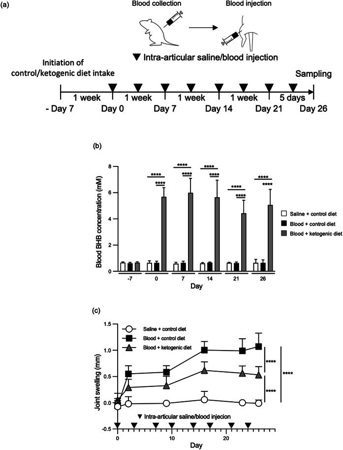
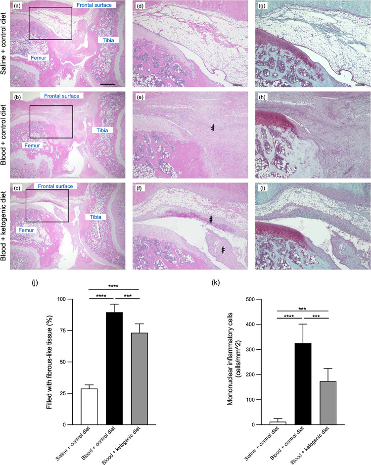
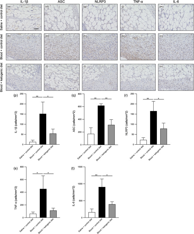
Arthropathy is a common complication in haemophilia and decreases quality of life. It has been known that concentrations of β-hydroxybutyrate (BHB) in blood are increased by a ketogenic diet, and elevated levels of circulating BHB restricts the progression of inflammation-mediated joint pathological changes. We hypothesized that elevation of blood BHB concentrations could be effective for reducing the progression of bleeding-induced arthropathy by moderating the inflammatory responses of macrophages. In this study, we investigated whether BHB alleviates the arthropathy caused by repeated intra-articular blood injection in rats. To increase blood BHB levels, rats were fed with ketogenic diet. Repeated intra-articular blood injection induced significant joint swelling, whereas ketogenic diet intake significantly increased blood BHB concentrations and ameliorated the joint swelling. The periarticular tissue-fibrosis observed in the control diet intake group appeared to be significantly alleviated in the ketogenic diet intake group. In addition, the IL-1β, which is involved in the progression of arthropathy, levels in the supernatants of blood-exposed macrophages derived from THP-1 cell line were significantly suppressed by BHB supplementation. In summary, BHB moderated the pathological joint changes caused by intra-articular blood exposure.


Conflict of interest statement

Declarations. Competing interests: RK, AS, KT, NM, SH, TS, YN, and MS: members of Medical Biology of Thrombosis and Hemostasis established by Nara Medical University and Chugai Pharmaceutical Co., Ltd.RK, NM, SH, TS, YN, and YY: employees of Chugai Pharmaceutical Co., Ltd.RK, NM, SH, and YY: stock ownership of Chugai Pharmaceutical Co., Ltd.TS, YN, YY, and MS: Patents for inventions relating to products of Chugai Pharmaceutical Co., Ltd.AS: speaker’s bureau from CSL Behring.KT: grants or research support from Japan Blood Products Organization, The Mother and Child Health Foundation and Novo Nordisk Pharma.SM, MT, and SK: no competing interest.MS: representative of Medical Biology of Thrombosis and Hemostasis collaborative research laboratory.; research support from Chugai Pharmaceutical Co., Ltd., Takeda Pharmaceutical Co., Ltd. and CSL Behring.; honoraria or consultation fees from Chugai Pharmaceutical Co., Ltd.; speaker’s bureau from Chugai Pharmaceutical Co., Ltd., CSL Behring, Sanofi, Bayer, Novo Nordisk Pharma, Takeda Pharmaceutical Co., Ltd., Pfizer and Fujimoto Seiyaku Corp. RK, NM, SH, TS, YN, and YY: employees of Chugai Pharmaceutical Co., Ltd. RK, NM, SH, and YY: stock ownership of Chugai Pharmaceutical Co., Ltd. TS, YN, YY, and MS: Patents for inventions relating to products of Chugai Pharmaceutical Co., Ltd. AS: speaker’s bureau from CSL Behring. KT: grants or research support from Japan Blood Products Organization, The Mother and Child Health Foundation and Novo Nordisk Pharma. SM, MT, and SK: no competing interest. MS: representative of Medical Biology of Thrombosis and Hemostasis collaborative research laboratory.; research support from Chugai Pharmaceutical Co., Ltd., Takeda Pharmaceutical Co., Ltd. and CSL Behring.; honoraria or consultation fees from Chugai Pharmaceutical Co., Ltd.; speaker’s bureau from Chugai Pharmaceutical Co., Ltd., CSL Behring, Sanofi, Bayer, Novo Nordisk Pharma, Takeda Pharmaceutical Co., Ltd., Pfizer and Fujimoto Seiyaku Corp.

Figures

Fig. 1
Fig. 1
Suppressive effects of ketogenic diet on joint swelling caused by repeated intra-articular blood injection. Control or ketogenic diet was commenced 7 days before the first intra-articular blood injection (Day 0). (a) Total of 8 intra-articular injections of autologous blood were performed twice a week from Day 0. Knee joints were assessed on Day 26. (b) β-hydroxybutyrate (BHB) concentrations in blood were measured on Days − 7, 0, 7, 14, 21, and 26. (c) Joint swelling was evaluated before and after the initiation of intra-articular injection of autologous blood. The days of blood injection are shown as black triangles (a and c). In the bar graph and line plot (b and c), white identifies the intra-articular saline injection + control diet (Saline + control diet) group, black denotes the intra-articular blood injection + control diet (Blood + control diet) group, and gray illustrates the intra-articular blood injection + ketogenic diet (Blood + ketogenic diet) group. Data are shown as mean and SD (n = 5–7). Intergroup comparisons were assessed by two-way ANOVA. Bonferroni multiple comparison test was used for comparisons among each group (****p < 0.0001).
Fig. 2
Fig. 2
Suppressive effects of ketogenic diet on arthrofibrosis and infiltration of mononuclear inflammatory cells after repeated intra-articular blood injections. The histology of knee joints on post intra-articular injection day 26 was compared, in the intra-articular saline injection + control diet (Saline + control diet) group (a, d, and g), the intra-articular blood injection + control diet (Blood + control diet) group (b, e, and h), and the intra-articular blood injection + ketogenic diet (Blood + ketogenic diet) group (c, f, and i) using haematoxylin-eosin and Safranin O-fast green staining. Representative low-power field images (a, b, and c), and selected high-power images of corresponding rectangular areas (d, e, f, g, h, and i) are illustrated. The scale bar in (a) indicates 1000 μm, and the scale bars in (d) and (g) indicate 200 μm. The frontal surface of the knee is located at the top in the images, the femur is on the left side, and tibia is at right side of the images. The percentage of fibrous-like tissue area (♯) in the infrapatellar fat pad (j) was measured for the evaluation of fibrosis using ImageJ software. The number of infiltrating inflammatory mononuclear cells per 1 mm2 in the periarticular tissues was counted (k). White: Saline + control diet group, black: Blood + control diet group, and gray: Blood + ketogenic diet group (j and k). Data are shown as mean and SD. Intergroup comparisons were assessed by one-way ANOVA. Bonferroni multiple comparison test was used for comparisons among each group (***p < 0.001, ****p < 0.0001).
Fig. 3
Fig. 3
Suppressive effects of ketogenic diet on the number of inflammatory cytokine-positive cells and inflammasome component protein-positive cells surrounding knee joint after repeated intra-articular blood injections. The immunohistochemical staining of knee joints on post intra-articular injection day 26 was compared, in the intra-articular saline injection + control diet (Saline + control diet) group (a, d, g, j, and m), the intra-articular blood injection + control diet (Blood + control diet) group (b, e, h, k, and n), and the intra-articular blood injection + ketogenic diet (Blood + ketogenic diet) group (c, f, i, l, and o) using anti-IL-1β antibody (ac), anti-ASC antibody (df), anti-NLRP3 antibody (gi), anti-TNF-α antibody (jl), and anti-IL-6 antibody (mo). The scale bar in (a) indicates 50 μm. The number of inflammatory cytokine-positive cells (IL-1β, TNF-α, and IL-6) and inflammasome component protein-positive cells (ASC and NLRP3) per 1 mm2 in the periarticular tissues was counted (pt). White: Saline + control diet group, black: Blood + control diet group, and gray: Blood + ketogenic diet group (pt). Data are shown as mean and SD. Intergroup comparisons were assessed by one-way ANOVA. Bonferroni multiple comparison test was used for comparisons among each group (*p < 0.05, **p < 0.01).
Fig. 4
Fig. 4
Direct effects of BHB on suppressing joint swelling caused by intra-articular blood injection. Rats fed with a chow diet were administered single intraperitoneal (i.p.) injection of PBS or BHB (8 mmol/kg) under isoflurane anaesthesia followed fifteen minutes by an intra-articular injection of whole blood. (a) Different groups of animals were assigned (n = 5–7 in each group) to receive autologous blood (Group 1 and 2), or blood with normal concentrations of BHB and with high concentrations of BHB (Group 3 and 4) as described in methods. (b) BHB concentrations were measured before the intra-articular blood injections. (c) Joint swelling was assessed two days after the blood injections. White: Group 1, black: Group 2, gray: Group 3, and light gray: Group 4 (a, b, and c). Data are shown as mean and SD. Intergroup comparisons were assessed by one-way ANOVA. Bonferroni multiple comparison test was used for comparisons among each group (*p < 0.05, **p < 0.01, ****p < 0.0001).
Fig. 5
Fig. 5
Direct effects of BHB on suppressing IL-1β excretion from macrophages differentiated from the THP-1 cell line. THP-1 cells were incubated in RPMI1640 medium containing 30 nM Phorbol 12-myristate 13-acetate (PMA) for 96 h to initiate macrophage differentiation. After induction, cells incubated in RPMI1640 medium with or without 1 ~ 10% human blood for 24 h prior to quantitation of IL-1β, TNF-α, and IL-6 by ELISA (a, b, and c). BHB (1 ~ 10 mM) or PBS was added to wells containing 10% blood (d, e, and f). Data are shown as mean and SD. Intergroup comparisons were assessed by one-way ANOVA. Bonferroni multiple comparison test was used for comparisons among each group (*p < 0.05, ****p < 0.0001).
Fig. 6
Fig. 6
Effects of BHB on the expression of IL-1β in macrophages differentiated from THP-1 cells. (a) THP-1-derived macrophages were incubated in RPMI1640 medium with or without 1 ~ 10% blood, and IL-1β mRNA expression was quantified by qPCR 24 h after blood addition. (b) BHB was added to wells containing 10% blood. Data are shown as mean and SD. Intergroup comparisons were assessed by one-way ANOVA. Bonferroni multiple comparison test was used for comparisons among each group (****p < 0.0001).

References

    1. Smolen, J. S., Aletaha, D. & McInnes, I. B. Rheumatoid arthritis. Lancet. 388, 2023–2038. 10.1016/s0140-6736(16)30173-8 (2016). - PubMed
    1. Goldring, M. B. & Otero, M. Inflammation in osteoarthritis. Curr. Opin. Rheumatol.23, 471–478. 10.1097/BOR.0b013e328349c2b1 (2011). - PMC - PubMed
    1. Evers, B. J. et al. Post-traumatic knee osteoarthritis: The role of inflammation and hemarthrosis on disease progression. Front. Med. (Lausanne). 9, 973870. 10.3389/fmed.2022.973870 (2022). - PMC - PubMed
    1. Goldberg, E. L. et al. β-Hydroxybutyrate deactivates Neutrophil NLRP3 Inflammasome to relieve gout flares. Cell. Rep.18, 2077–2087. 10.1016/j.celrep.2017.02.004 (2017). - PMC - PubMed
    1. Kong, G., Wang, J., Li, R., Huang, Z. & Wang, L. Ketogenic diet ameliorates inflammation by inhibiting the NLRP3 inflammasome in osteoarthritis. Arthritis Res. Ther.24, 113. 10.1186/s13075-022-02802-0 (2022). - PMC - PubMed

LinkOut - more resources