. 2024 Nov 30;21(1):312.
doi: 10.1186/s12974-024-03283-5.

Sitagliptin eye drops prevent the impairment of retinal neurovascular unit in the new Trpv2+/- rat model

Affiliations

Sitagliptin eye drops prevent the impairment of retinal neurovascular unit in the new Trpv2+/- rat model

Hugo Ramos et al. J Neuroinflammation. .

Abstract

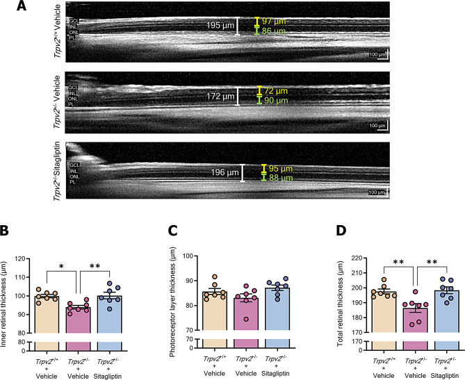
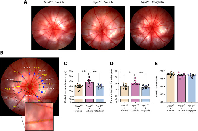
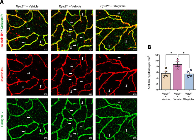
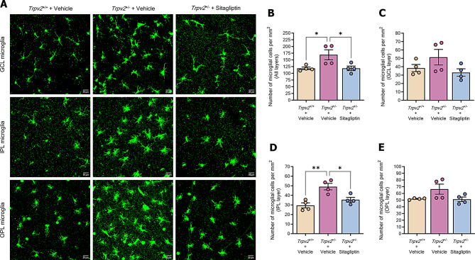
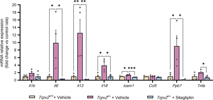
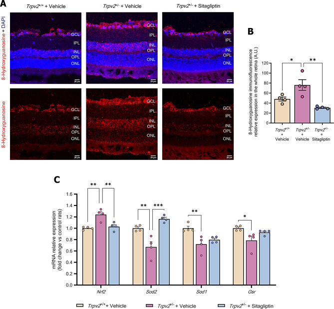
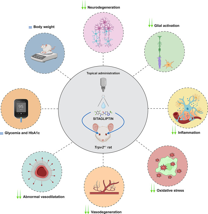
Impaired function of the retinal neurovascular unit (NVU) is an early event in diabetic retinopathy (DR). It has been previously shown that topical delivery of the dipeptidyl peptidase-4 (DPP-4) inhibitor sitagliptin can protect against diabetes-mediated dysfunction of the retinal NVU in the db/db mouse. The aim of the present study was to examine whether sitagliptin could prevent the DR-like lesions within the NVU of the new non-diabetic model of DR, the Trpv2 knockout rat (Trpv2+/-). For that purpose, at 3 months of age, Trpv2+/- rats were topically treated twice daily for two weeks with sitagliptin or PBS-vehicle eyedrops. Trpv2+/+ rats treated with vehicle served as the control group. Body weight and glycemia were monitored. Optical coherence tomography recordings, fundus images and retinal samples were obtained to evaluate sitagliptin effects. The results revealed that sitagliptin eye drops had no effect on body weight or glycemia. Vehicle-treated Trpv2+/- rats exhibited retinal thinning and larger diameters of major retinal blood vessels, upregulation of inflammatory factors and oxidative markers, glial activation and formation of acellular capillaries. However, topical administration of sitagliptin significantly prevented all these abnormalities. In conclusion, sitagliptin eye drops exert a protective effect against DR-like lesions in Trpv2+/- rats. Our results suggest that sitagliptin eye drops carry significant potential to treat not only early-stages of DR but also other diseases with impairment of the NVU unrelated to diabetes.

Keywords: DPP-4 inhibitor; Diabetes; Diabetic retinopathy; Sitagliptin; Trpv2.


Conflict of interest statement

Declarations. Ethical approval: Animal experiments were approved by the Animal Welfare Ethical Review Body (AWERB) of Queen’s University Belfast. The protocol complied with the UK Home Office Animals (Scientific Procedures) Act 1986 (project license, PPL2888) and the Association for Research in Vision and Ophthalmology (ARVO) statement for the use of animals in ophthalmology and vision research. Competing interests: Two of the authors (Cristina Hernández and Rafael Simó) are inventors of the patent PCT/EP2017/060234, which is related to use of dipeptidyl peptidase-4 inhibitors (sitagliptin) for topical eye treatment of retinal neurodegenerative diseases.

Figures

Fig. 1
Fig. 1
Evaluation of physiological parameters. A,B Body weight A glycaemic B measurements during the experimental course of Trpv2+/− rats treated with sitagliptin (blue) or vehicle (purple) and Trpv2+/+ wild type rats treated with vehicle (cream-colour); n = 7. C Bar graph illustrating the average HbA1c measurements per each group at the end of the experiment, prior to euthanasia. Control homozygous rats are represented with a green bar, while vehicle-treated heterozygous rats are represented with a red bar and sitagliptin-treated heterozygous rats with a blue bar; n = 7. D Bar graph showing mean values from the RT-qPCR analysis of Trpv2 gene in vehicle-treated Trpv2+/+ rats (cream-colour) and in Trpv2+/− heterozygous rats that received vehicle (purple) or sitagliptin eye drops (blue). Results are presented as fold change vs. control mice; n = 4. ns = no significance, ** p < 0.01
Fig. 2
Fig. 2
Retinal thickness assessment through SD-OCT. Measurements were obtained at a consistent eccentricity of 1500 μm from the optic disc in four quadrants (superior, inferior, nasal, and temporal) of the eye. Total retinal thickness, inner retinal thickness, and photoreceptor layer thickness were measured in each quadrant, and the final value was calculated as the mean across all four quadrants. A Representative images of SD-OCT thickness measurements of inner retina (yellow bar), photoreceptor layer (green bar), and total neuroretina from Trpv2+/− rats treated with vehicle or sitagliptin and wild-type rats treated with vehicle. Scale bar: 100 μm. B-D Bar graphs showing the mean values for the thickness measurements of inner retina B, photoreceptor layer C and total retina D. Control homozygous rats are represented with a cream-colored bar, while vehicle-treated heterozygous rats are represented with a purple bar and sitagliptin-treated heterozygous rats with a blue bar; n = 7. * p < 0.05, ** p < 0.01
Fig. 3
Fig. 3
Funduscopic examination and diameter measurements of major retinal vessels. Fundus images were captured with the optic nerve at the centre and analysed using ARIA software, with the optic disc manually defined. Arteriolar and venular diameters were measured in a predefined region located at a distance ranging from one-and-a-half to two-and-a-half disc diameters from the optic disc. The four largest arterioles and venules were selected for consistent analysis, and data from both eyes were averaged per animal for statistical evaluation. A Representative fundus images of all the studied experimental groups. B Exemplifying images of the procedure of diameter analysis for major veins and arteries of the retina. The small green circle represents the optic disc, while the medium and large green circles delimit the studied area for all vessels. The yellow labelling illustrates the vessel under analysis at that time, while the enlarged image shows each of the measurements with thin yellow lines. C-E Bar graphs depicting the mean values of diameter measurements for main retinal veins C and arteries D and arterio-venous ratio E among experimental groups. Control homozygous rats are represented with cream-colored bars, while vehicle-treated heterozygous rats are represented with purple bars and sitagliptin-treated heterozygous rats with blue bars; n = 7. * p < 0.05, ** p < 0.01
Fig. 4
Fig. 4
Analysis of acellular capillary formation. A Representative confocal Z-stack images of retinal whole mount preparations stained with collagen IV (green) and isolectin-B4 (red) focused at the superficial vascular plexus. Acellular capillaries, indicated with white arrows, are identifiable as deteriorated vascular segments showing positivity for collagen IV but negativity for isolectin-B4. Scale bars: 20 μm. B Bar graph illustrating the average number of acellular capillaries per experimental group. Control homozygous rats are represented with a cream-colored bar, while heterozygous rats treated with vehicle or sitagliptin are represented with a purple bar and a blue bar respectively. n = 4. * p < 0.05
Fig. 5
Fig. 5
Evaluation of Müller cell activation. A Representative confocal Z-stack images of retinal criosections stained with GFAP (green) and DAPI (blue) from each experimental group. Scale bars: 20 μm. B Bar graph with the average numbers of GFAP + fibres located across the whole retina. Control Trpv2+/+ rats are represented with cream-colored bars, while vehicle-treated Trpv2+/− rats are represented with red bars and sitagliptin-treated Trpv2+/− rats with blue bars; n = 4. * p < 0.05, ** p < 0.01. C-F Bar graphs illustrating mean values from the RT-qPCR analysis of Kncj10 C, Aqp4 D, Glul E and Vegfa F genes in vehicle-treated Trpv2+/+ homozygous rats (cream-colour) and in Trpv2+/− heterozygous rats that received vehicle (purple) or sitagliptin eye drops (blue). Results are presented as fold change vs. control mice; n = 4. GCL (ganglion cell layer), IPL (inner plexiform layer), INL (inner nuclear layer), OPL (outer plexiform layer), ONL (outer nuclear layer). * p < 0.05, ** p < 0.01
Fig. 6
Fig. 6
Assessment of number of microglial cells. A Representative confocal Z-stack images of whole-mount preparations labelled with IBA-1 (green) from each experimental group. Scale bars: 20 μm. B-E Bar graphs depicting the average number of microglial cells per mm2 of GCL B, IPL c, OPL D and all E)microglial layers of the retina for each studied experimental group. Control homozygous rats are represented with a cream-colored bar, while heterozygous rats treated with vehicle or sitagliptin are represented with a purple bar and a blue bar respectively. n = 4. GCL (ganglion cell layer), IPL (inner plexiform layer), OPL (outer plexiform layer). * p < 0.05, ** p < 0.01
Fig. 7
Fig. 7
Evaluation of the pro-inflammatory environment. A Bar graph showing the mean values from the RT-qPCR analysis of genes that codify for some pro-inflammatory cytokines in vehicle-treated Trpv2+/+ homozygous rats (cream-colour) and in Trpv2+/− heterozygous rats that received vehicle (purple) or sitagliptin eye drops (blue). Results are presented as fold change vs. control mice; n = 4. * p < 0.05, ** p < 0.01, *** p < 0.001
Fig. 8
Fig. 8
Examination of DNA oxidative damage and retinal antioxidant mechanisms. A Representative confocal Z-stack images of 8-hydroxyguanosine (red) and DAPI (blue) stainings on retinal criosections. B Bar graph illustrating the mean values of 8-hydroxyguanosine immunofluorescence intensity of each experimental group. Control homozygous rats are represented with cream-colored bars, while vehicle-treated heterozygous rats are represented with purple bars and sitagliptin-treated heterozygous rats with blue bars; n = 4. c Bar graph illustrating mean values from the RT-qPCR analysis of Nrf2, Sod1, Sod2 and Gsr genes in vehicle-treated Trpv2+/+ homozygous rats (cream-colour) and in Trpv2+/− heterozygous rats that received vehicle (purple) or sitagliptin eye drops (blue). Results are presented as fold change vs. control mice; n = 4. GCL (ganglion cell layer), IPL (inner plexiform layer), INL (inner nuclear layer), OPL (outer plexiform layer), ONL (outer nuclear layer). * p < 0.05, ** p < 0.01, *** p < 0.001
Fig. 9
Fig. 9
Primary effects of sitagliptin eye drops in the Trpv2+/− model after two-weeks of twice-daily administration. Topical application of sitagliptin resulted in a reduction of neurodegeneration (preservation of total and inner retinal thickness), glial activation (reduction in GFAP-positive fibers and microglial cells), inflammation (decreased pro-inflammatory cytokines), oxidative stress (diminished DNA/RNA damage and maintenance of antioxidant elements), vasodegeneration (protection against the formation of acellular capillaries) and abnormal vasodilation. These effects were independent of changes in body weight, glycemia, and HbA1c levels

References

    1. Wong TY, Cheung CM, Larsen M, Sharma S, Simó R. Diabetic retinopathy. Nat Rev Dis Primers. 2016;2:16012. 10.1038/nrdp.2016.12. - PubMed
    1. Duh EJ, Sun JK, Stitt AW. Diabetic retinopathy: current understanding, mechanisms, and treatment strategies. JCI Insight. 2017;2(14):e93751. 10.1172/jci.insight.93751. - PMC - PubMed
    1. Simó R, Stitt AW, Gardner TW. Neurodegeneration in diabetic retinopathy: does it really matter? Diabetologia. 2018;61(9):1902–12. 10.1007/s00125-018-4692-1. - PMC - PubMed
    1. Simó R, Hernández C. What else can we do to prevent diabetic retinopathy? Diabetologia. 2023;66(9):1614–21. 10.1007/s00125-023-05940-5. - PMC - PubMed
    1. Hernández C, Bogdanov P, Corraliza L, et al. Topical administration of GLP-1 receptor agonists prevents retinal neurodegeneration in experimental diabetes. Diabetes. 2016;65(1):172–87. 10.2337/db15-0443. - PubMed

MeSH terms

Substances

LinkOut - more resources