. 2025 Jul;35(4):e13322.
doi: 10.1111/bpa.13322. Epub 2024 Dec 1.

Neuropathological and cerebrospinal fluid correlates of choroid plexus inflammation in progressive multiple sclerosis

Affiliations

Neuropathological and cerebrospinal fluid correlates of choroid plexus inflammation in progressive multiple sclerosis

R Magliozzi et al. Brain Pathol. 2025 Jul.

Abstract

Among the intrathecal inflammatory niches where compartmentalized inflammation persists and plays a pivotal role in progressive multiple sclerosis (MS), choroid plexus (CP) has recently received renewed attention. To better characterize the neuropathological/molecular correlates of CP in progressive MS and its potential link with other brain inflammatory compartments, such as perivascular spaces and leptomeninges, the levels, composition and phenotype of CP immune infiltration in lateral ventricles of the hippocampus were examined in 40 post-mortem pathologically confirmed MS and 10 healthy donors, using immunochemistry/immunofluorescence and in-situ sequencing. Significant inflammation was detected in the CP of 21 out of the 40 MS cases (52%). The degree of CP inflammation was found correlated with: number of CP macrophages (R: 0.878, p = 1.012 x 10⁻¹³) and high frequency of innate immune cells expressing the markers MHC-class II, CD163, CD209, CD11c, TREM2 and TSPO; perivascular inflammation (R: 0.509, p = 7.921 x 10⁻⁴), and less with meningeal inflammation (R: 0.365, p = 0.021); number of active lesions (R: 0.51, p: 3.524 x 10⁻⁵). However, it did not significantly correlate with any clinical/demographic characteristics of the examined population. In-situ sequencing analysis of gene expression in the CP of 3 representative MS cases and 3 controls revealed regulation of inflammatory pathways mainly related to 'type 2 immune response', 'defense to infections', 'antigen processing/presentation'. Analysis of 78 inflammatory molecules in paired post-mortem CSF, the levels of fibrinogen (R: 0.640, p = 8.752 x 10⁻⁶), PDGF-bb (R: 0.470, p = 0.002), CXCL13 (R: 0.428, p = 0.006) and IL15 (R: 0.327, p = 0.040) were correlated with extent of CP inflammation. Elevated fibrinogen and complement deposition were found in CP and in underlying subependymal periventricular areas, according to "surface-in" gradient associated with concomitant prominent microglia activation. CP inflammation, predominantly characterized by innate immunity, represents another key determinant of intrathecal, compartmentalised inflammation persisting in progressive MS, which may be possibly activated by fibrinogen and influence periventricular pathology, even without substantial association with clinical features.

Keywords: choroid plexus; gradient; inflammation; multiple sclerosis.


Conflict of interest statement

The authors declare no conflicts of interest related to this study.

Figures

FIGURE 1
FIGURE 1
Semi‐quantitative scoring of CP (indicated by asterisk *) inflammation was performed in the lateral ventricle included in the hippocampus (A–C) of the examined MS cases. By summing the number of CD3+ T cells (D–F), CD20+ B cells (G–I) and CD68+ cells (J–L) for each examined patients the following scores were proposed: Score 0 corresponds to absent/scarce (0–5 cells), 1 corresponds to moderate (6–25 cells), 2 corresponds to high (26–50 cells) inflammatory infiltrate. Scale bars: 1000 μm (A–C), 100 μm (D–L).
FIGURE 2
FIGURE 2
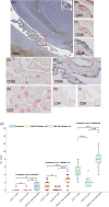
Organized lymphocyte aggregates were never detected in the CP. However, in some MS cases, characterized by elevated meningeal inflammation, small compact cell aggregates, CD3+, CD20+, and CD68+ cells, were identified along the “tela coroidea” (square in A and magnifications B–D), while the CP infiltrating cells were mainly found scattered in the stroma. Cell count analysis (E–J) performed comparing 10 healthy donors (light blue), MS with CP = 0 (yellow) and MS with CP >0 (green) demonstrated: significant increase of T‐cells (vs. MS CP = 0, p: 1.261 × 10−5; vs. MS CP >0, p: 8.136 × 10−6), B‐cells (vs. MS CP = 0, p: 0.021; vs. MS CP >0, p: 1.094 × 10−5), and macrophages (vs. MS CP = 0, p: 1.178 × 10−5; vs. MS CP >0, p: 1.029 × 10−5). In addition, the presence of inflammation within the CP was coupled with significant increase of all the type of infiltrating cells (MS CP >0) compared to MS patients characterized by lack of inflammation (MS CP = 0) (T cells p: 3.564 × 10−7; B cells, p: 2.060 × 10−6; macrophages, p: 1.006 × 10−7). Among the CD3+ T cells infiltrating the CP, preponderance of CD4+ T cells was observed (G–I). Scale bars: 500 μm (A), 100 μm (B–I).
FIGURE 3
FIGURE 3
Neuropathology characterization of CP innate immune infiltration. Frequent MHCII+ activated macrophages (A), occasionally including intracytoplasmic LFB myelin debris (inset in A and in B), were observed in the inflamed CP (in MS CP >0), mainly in the stromal space between the epithelial and endothelial layers (stromal macrophages). Macrophage CP activity was further characterized by elevated expression of haptoglobin/hemoglobin receptor, CD163 (B), of the dendric cell markers CD209/DCsign (Figure 3C–F, indicated by arrows) and of CD11c, marker of antigen presentation CD11c (G, H, indicated by arrows). Scattered CP infiltrating cells expressing the activation myeloid markers TREM2, sporadically with endothelial morphology and localization (I–J, indicated by arrows) were observed. Only sporadically CD16+ (K, indicated by arrows) or CD66b+ (L, indicated by arrows) granulocytes were detected, predominantly within the CP blood vessels. Scale bars: 100 μm (A–F), 50 μm (G–L).
FIGURE 4
FIGURE 4
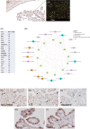
In‐situ sequencing (Cartana) of 157 inflammatory genes on the CP of a representative MS case (A, B). List of the 19 genes significant overexpressed (fold change >1.5, p < 0.05) in the CP of MS compared to healthy donors (C). Subnetwork analysis of the top 12 genes (fold change >2, p < 0.05) (D) extensively explained in Figure S2. Immunohistochemistry validation of some of the significantly deregulated genes found by ISS Cartana analysis, confirmed the presence in the CP on serial brain sections of scattered cells expressing SELL/CD62L+ (E) and RASGRP2 (G), and of frequent cells expressing IL15 (F) or TSPO (H, I). Scale bars: 200 μm (A, B), 100 μm (E–G), 50 μm (G–L).
FIGURE 5
FIGURE 5
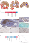
Graphical representation of the widespread white matter (blue color mask) and grey matter (cortical and deep grey matter; red color mask) demyelination in MS cases characterized by substantial inflammation of the CP (A). The presence of lesions was mainly associated with periventricular white matter and hippocampus (A). Spearman correlation analysis (B) between CP inflammation and the other quantified neuropathological parameters shows significant positive correlation (darker red) between degree of CP inflammation and degree of perivascular inflammation (R: 0.509, p: 7.921 × 10−4), with meningeal inflammation (R: 0.365, p: 0.021) and with the number of active lesions (R: 0.51, p: 3.524 × 10−5) identified in the surrounding periventricular areas (B). Extensive subependymal active demyelination (C) with intense microglia activation, characterized by expression of MHC‐class II, TMEM119, CD163, and iNOs (D–H) were detected in the hippocampus. Spearman correlation analysis between degree of CP inflammation and clinical features of the examined MS cases did not reveal any significant correlation (I). Survival analysis that compared the populations with (red line) and without (blue line) CP inflammation, with respect to the age at death did not found significantly difference (p: 0.391), even if a higher percentage of subjects without CP inflammation were characterized by later age at death (J). Scale bars: 500 μm (C), 200 μm (D), 100 μm (E–I).
FIGURE 6
FIGURE 6
Spearman correlation analysis (A) between CP inflammation and Among the 78 molecules examined in the CSF of the same MS cases: Degree of CP inflammation was found significantly and positively correlated (darker red and p values in bold) only with 4 markers: Fibrinogen (R: 0.640, p: 8.752 × 10−6), PDGF‐bb (R: 0.470, p: 0.002), CXCL13 (R: 0.428, p: 0.006) and IL15 (R: 0.327, p: 0.040) (A). CSF fibrinogen levels were significantly different not only in MS cases compared to controls (B; p: 0.0001), but also between the two populations (p: 8.654 × 10−6) with and without CP inflammation (C). Neuropathological analyzing revealed fibrinogen immunoreactivity in active subpial cortical lesions (D–F) or sub‐ependymal lesions (G–I) in the hippocampus of the examined MS cases, mainly in cells morphologically resembling microglia, astrocytes or neuronal cells (J, K), as validated by double immunofluorescence (L–N). Schematic diagram of assessment of gradient of fibrinogen deposition from CSF surfaces towards inner brain regions, either at the pial or at the ependymal surface (O) according to the potential presence of a “surface‐in” gradient in 10 of the examined MS cases characterized by the presence of at least one subpial cortical lesion and one subependymal one. Sub‐pial (P, Q) and sub‐ependyma (R, S) gradients of fibrinogen+ cells increased density in the external areas close to CSF/brain interface. Most of the fibrinogen+ cells were identified as MHC+ microglia (Q, 59% cortical layer I in subpial lesions; S, 53% field I in subpendymal lesions), in particular in the most external areas, while less abundant proportion of MAP2+ neurons and GFAP+ astrocytes were identified in the same regions (Q, S). Scale bars: 1000 μm (O), 100 μm (D–K), 50 μm (L–N).

References

    1. Kaur C, Rathnasamy G, Ling E‐A. The choroid plexus in healthy and diseased brain. J Neuropathol Exp Neurol. 2016;75:198–213. 10.1093/jnen/nlv030 - DOI - PubMed
    1. Kivisäkk P, Mahad DJ, Callahan MK, Trebst C, Tucky B, Wei T, et al. Human cerebrospinal fluid central memory CD4+ T cells: evidence for trafficking through choroid plexus and meninges via P‐selectin. Proc Natl Acad Sci. 2003;100:8389–8394. 10.1073/pnas.1433000100 - DOI - PMC - PubMed
    1. Monaco S, Nicholas R, Reynolds R, Magliozzi R. Intrathecal inflammation in progressive multiple sclerosis. Int J Mol Sci. 2020;21:8217. 10.3390/ijms21218217 - DOI - PMC - PubMed
    1. Vercellino M, Votta B, Condello C, Piacentino C, Romagnolo A, Merola A, et al. Involvement of the choroid plexus in multiple sclerosis autoimmune inflammation: a neuropathological study. J Neuroimmunol. 2008;199:133–141. 10.1016/j.jneuroim.2008.04.035 - DOI - PubMed
    1. Rodríguez‐Lorenzo S, Konings J, van der Pol S, Kamermans A, Amor S, van Horssen J, et al. Inflammation of the choroid plexus in progressive multiple sclerosis: accumulation of granulocytes and T cells. Acta Neuropathol Commun. 2020;8:9. 10.1186/s40478-020-0885-1 - DOI - PMC - PubMed