. 2024 Dec 1;15(1):10450.
doi: 10.1038/s41467-024-54635-x.

AAV delivery strategy with mechanical support for safe and efficacious cardiac gene transfer in swine

Affiliations

AAV delivery strategy with mechanical support for safe and efficacious cardiac gene transfer in swine

Renata Mazurek et al. Nat Commun. .

Abstract

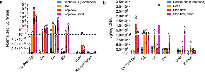
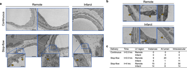
Adeno-associated virus-based gene therapy is a promising avenue in heart failure treatment, but has shown limited cardiac virus uptake in humans, requiring new approaches for clinical translation. Using a Yorkshire swine ischemic heart failure model, we demonstrate significant improvement in gene uptake with temporary coronary occlusions assisted by mechanical circulatory support. We first show that mechanical support during coronary artery occlusions prevents hemodynamic deterioration (n = 5 female). Subsequent experiments show that coronary artery occlusions during gene delivery improve gene transduction, while adding coronary sinus occlusion (Stop-flow) further improves gene expression up to >1 million-fold relative to conventional intracoronary infusion. Complete survival during and after delivery (n = 10 female, n = 10 male) further indicates safety of the approach. Improved cardiac gene expression correlates with virus uptake without an increase in extra-cardiac expression. Stop-flow delivery of virus-sized gold nanoparticles exhibits enhanced endothelial adherence and uptake, suggesting a mechanism independent of virus biology. Together, utilizing mechanical support for cardiac gene delivery offers a clinically-applicable strategy for heart failure-targeted therapies.


Conflict of interest statement

Competing interests: Part of this study was supported by a research grant from Abiomed, Inc. to the institution. K.I. serves as the principal investigator on the grant from Abiomed, Inc. K.I. received an honorarium from Abiomed, Inc. and served as a consultant for Pfizer, Inc., and Gordian Biotechnology. T.S. and T.K. were supported by A-CURE Research Fellowship supported by Abiomed, Inc.. K.I. and R.J.H. have a patent, “SYSTEMS AND METHODS FOR LEFT VENTRICULAR UNLOADING IN BIOLOGIC THERAPY OR VECTORED GENE THERAPY” Publication number: 20200305888. All other authors declare no competing interests.

Figures

Fig. 1
Fig. 1. Overall study design.
Acute myocardial infarction was induced in Yorkshire pigs by temporal balloon occlusion of the left anterior descending artery (LAD) at day 0. One week later, animals were used for acute hemodynamic study (n = 5), acute nanoparticle study (n = 3), and AAV gene delivery study (n = 20), where n are independent animals and experimental procedures. AAV was injected via intracoronary delivery, followed by tissue harvest at 5 weeks for expression analysis. Intracoronary AAV injection was performed by one of five delivery methods: Continuous without mechanical circulatory support (MCS) (Continuous), Continuous with MCS (Continuous, MCS), Coronary artery occlusion with MCS (CAO), Stop-flow with MCS (Stop-flow), or Stop-flow without MCS and short occlusions (Stop-flow, short). AAV was delivered through the balloon wire lumen during balloon occlusions in the LAD (90 s each) and left circumflex artery (60 s each) in CAO and Stop-flow groups and for 15 s each in the same arteries in the short Stop-flow group.
Fig. 2
Fig. 2. Mechanical circulatory support provided hemodynamic stability during coronary balloon occlusions.
a Blood pressure was monitored in five animals one week after myocardial infarction (MI) during coronary artery balloon occlusions without and with mechanical cardiac support (MCS). b, c Following ischemic preconditioning, infarcted (LAD; b) and non-infarcted (LCx; c) coronary arteries were occluded with an over-the-wire coronary balloon for 5 min, and systolic aortic pressure was recorded at 0 and at 5 min. Source data are provided as a Source Data file.
Fig. 3
Fig. 3. Coronary artery occlusion improves cardiac gene expression.
a Different areas of myocardium from LV base, mid, and apex were collected to study gene distribution. Luciferase expression data were normalized to total protein. b, c Luciferase expression in the LV was compared between Continuous (n = 3) and CAO (n = 3) delivery groups (b) and between Continuous delivery without (n = 3) and with (n = 3) the addition of MCS (c) to assess the contribution of balloon occlusions or MCS to gene expression. All n-values represent biological replicates, with each point averaged from technical triplicates within the same animal. Expression is shown as mean ± SEM of biological replicates. Comparisons were conducted as a two-tailed t test with p-value set at 0.05. Source data are provided as a Source Data file. Dotted lines separate linear and logarithmic scales in (b). Infarct refers to the mid-anterior region. Border refers to the infarct-border at the mid-lateral region. Post, posterior; Epi, epicardial; Endo, endocardial; Lat, lateral; Sept, Septum.
Fig. 4
Fig. 4. Stop-flow delivery significantly increases cardiac gene expression.
Average normalized luciferase expression was compared between Stop-flow and other groups shown in Fig. 3. Continuous delivery groups with and without mechanical cardiac support were combined due to similar expression between the two. Dotted lines separate linear and logarithmic scales. Statistical significance is denoted at *p < 0.05. Continuous (Combined), n = 7; CAO, n = 3; Stop-flow, n = 4; Stop-flow, short, n = 3; IM, n = 3. All n-values represent biological replicates, with each point averaged from technical triplicates within the same animal. Expression is shown as mean ± SEM of biological replicates. Post, posterior; Epi, epicardial; Endo, endocardial; Lat, lateral; Sept, Septum. Source data are provided as a Source Data file. Statistics reflect comparisons by Kruskal-Wallis test where H, η2, p (exact): Apex (4.245, 0.10, 0.2498); Infarct (4.089, 0.08, 0.2645); Border (7.221, 0.32, 0.0471); Posterior endocardial (7.745, 0.37, 0.0322); Posterior epicardial (9.924, 0.53, 0.0051); Septum (5.681, 0.21, 0.1210); Base lateral (7.594, 0.35, 0.0361); Base septum (7.694, 0.36, 0.0336).
Fig. 5
Fig. 5. Stop-flow delivery increases AAV uptake and is associated with higher gene expression.
a Viral genome (vg) quantification by qPCR compared AAV uptake in LV regions after coronary balloon occlusions (CAO, n = 3; Stop-flow, n = 4; Stop-flow, short, n = 3) vs Continuous delivery (n = 7). The dotted line separates linear and logarithmic scales. b Vg in LV tissue regions was plotted against normalized luciferase in each delivery group. The dashed lines represent the average limits of detection for vg and luciferase expression. All n-values represent biological replicates, with each point averaged from technical triplicates within the same animal. Expression is shown as mean ± SEM of biological replicates. Post, posterior; Epi, epicardial; Endo, endocardial; Lat, lateral; Sept, Septum. Source data are provided as a Source Data file. Statistical significance is denoted at *p < 0.05. Statistics reflect delivery comparisons in vg by Kruskal-Wallis test where H, η2, p (exact): Apex (6.543, 0.27, 0.707); Infarct (2.865, − 0.01, 0.4351); Border (11.12, 0.62, 0.0017); Posterior endocardial (5.361, 0.18, 0.1142); Posterior epicardial (12.81, 0.75, 0.0002); Septum (0.2506, − 0.21, 0.9810); Base lateral (8.187, 0.40, 0.0239); Base septum (1.274, − 0.13, > 0.9999).
Fig. 6
Fig. 6. Stop-flow delivery allows for uptake in cardiomyocytes.
Examination of tissue distribution of vector via luciferase transgene probe (green) using RNAscope shows cardiomyocyte cytoplasmic distribution (arrowheads) in both posterior (remote) and infarct tissues. Sample areas of positive signal distribution are shown from posterior epicardial tissue and limited expression in infarct tissue (n = 2 biological replicates with highest luciferase expression). Acquisition at 40x magnification. Scale bar = 50 µm.
Fig. 7
Fig. 7. Intracoronary injection methods exhibit limited off-target gene expression.
a, b Average (a) luciferase expression and (b) AAV uptake in other cardiac and non-cardiac tissues in intracoronary delivery groups. Tissues that showed positive expression in any of the animals are shown. The dotted line in (a) separates linear and logarithmic scales. Continuous (Combined), n = 7; CAO, n = 3; Stop-flow, n = 4; Stop-flow, short, n = 3. All n-values represent biological replicates, with each point averaged from technical triplicates within the same animal. Expression is shown as mean ± SEM of biological replicates. Post, posterior; Epi, epicardial; CA, coronary artery; RV, right ventricle; LA, left atrium. Source data are provided as a Source Data file.
Fig. 8
Fig. 8. Stop-flow enhances AAV-sized gold nanoparticle uptake.
a, b Images are representative of gold nanoparticle detection (a) 30 min after Continuous vs Stop-flow delivery and (b) 4 h after Stop-flow delivery in remote (mid-posterior) and infarct (mid-anterior) tissues. Gold nanoparticles were not detected after Continuous delivery but were found with Stop-flow delivery (gold arrows), either attached at the endothelium-lumen interface or in vesicular structures. Images acquired at 4000x-6000x direct magnification were fitted to the same scale. Scale bar in (a), larger images = 600 nm; scale bar in (a) projections, (b) = 200 nm. c Observed instances of gold nanoparticle detection over equivalent grid areas and imaging time were recorded in each condition (n = 1 animal per condition, with duplicate tissues from each tissue region) representing gold nanoparticle distribution.

References

    1. Tsao, C. W. et al. Heart disease and stroke statistics-2023 update: A report from the American heart association. Circulation147, e93–e621 (2023). - PubMed
    1. Ravichandran, A. J., Romeo, F. J., Mazurek, R. & Ishikawa, K. Barriers in heart failure gene therapy and approaches to overcome them. Heart Lung Circ.32, 780–789 (2023). - PMC - PubMed
    1. Drouin, L. M. & Agbandje-McKenna, M. Adeno-associated virus structural biology as a tool in vector development. Future Virol.8, 1183–1199 (2013). - PMC - PubMed
    1. Hajjar, R. J. et al. Modulation of ventricular function through gene transfer in vivo. Proc. Natl. Acad. Sci. USA95, 5251–5256 (1998). - PMC - PubMed
    1. Hadri, L. et al. SERCA2a gene transfer enhances eNOS expression and activity in endothelial cells. Mol. Ther.18, 1284–1292 (2010). - PMC - PubMed

Publication types

LinkOut - more resources