. 2024 Dec 1;21(1):315.
doi: 10.1186/s12974-024-03292-4.

TRPM7 contributes to pyroptosis and its involvement in status epilepticus

Affiliations

TRPM7 contributes to pyroptosis and its involvement in status epilepticus

Xin Tong et al. J Neuroinflammation. .

Abstract

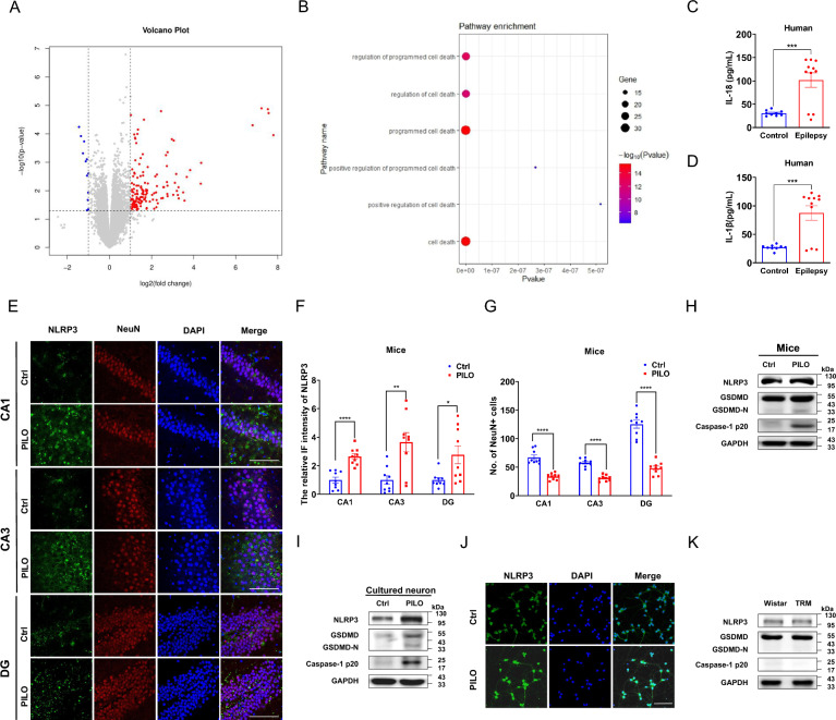
Background: Pyroptosis, a novel form of programmed cell death, has been implicated in neurodegeneration diseases. However, its role in status epilepticus (SE)-a condition characterized by prolonged or repeated seizures-remains inadequately understood.

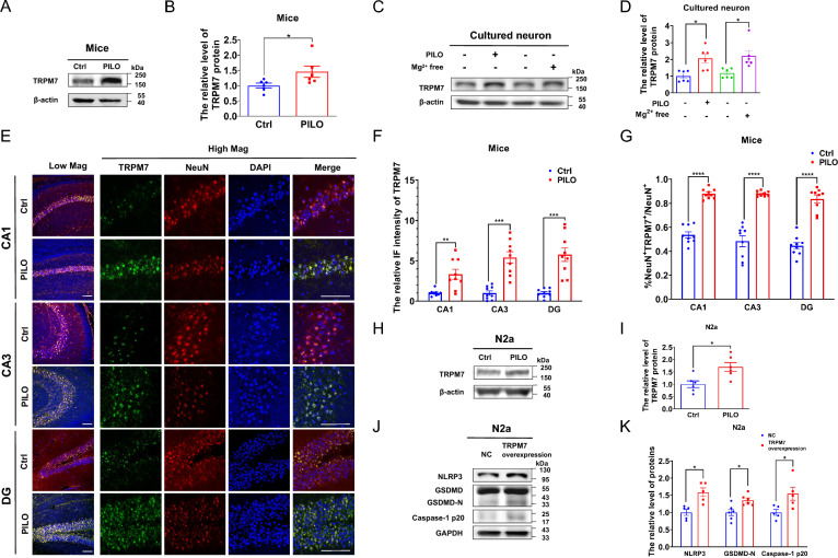
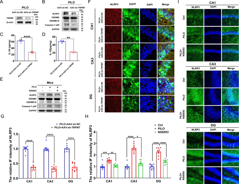
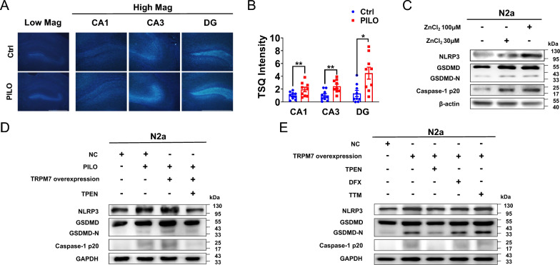
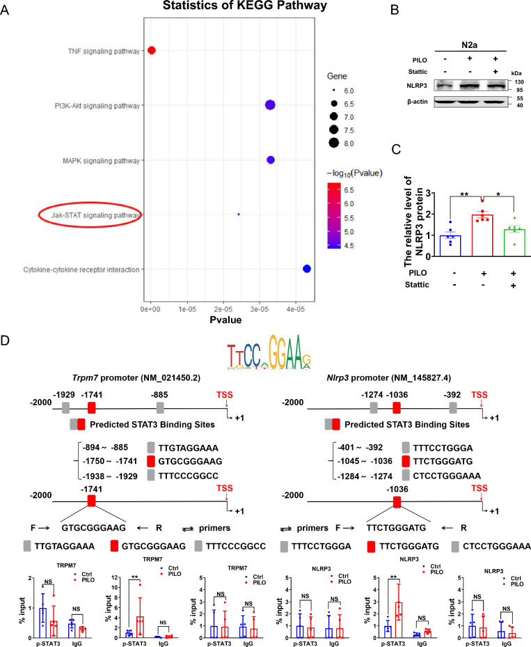
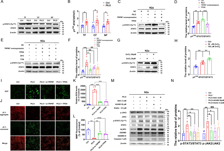
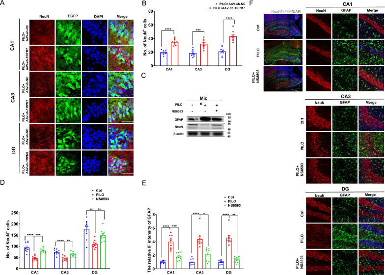
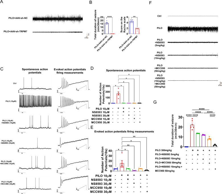
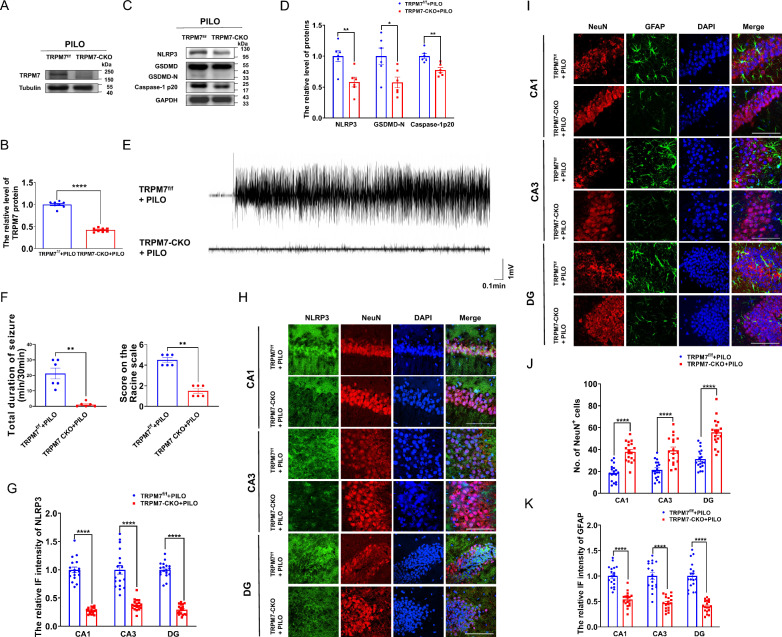
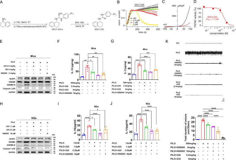
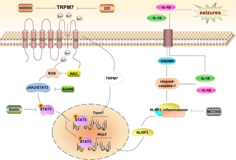
Methods: SE were induced by intraperitoneal injection of pilocarpine (PILO). Neuronal excitability was assessed through electroencephalogram (EEG) recordings and patch clamp. Chromatin immunoprecipitation (ChIP) assay was applied to verify the interaction of phosphorylated signal transducer and activator of transcription 3 (p-STAT3) protein with the promoters of Nlrp3 (the gene encoding NOD-like receptor family pyrin domain containing 3) and Trpm7 (transient receptor potential melastatin 7). To further investigate the role of TRPM7 in SE, AAV-sh-TRPM7-EGFP transfected mice and TRPM7 conditional knockout (TRPM7-CKO) mice were utilized.

Results: Our findings revealed elevated levels of IL-18 and IL-1β levels in primary epilepsy patients, along with increased expression level of the TRPM7 in SE models. Knockdown of TRPM7 alleviated neuronal damage and pyroptosis, reversing PILO-treated neuronal hyperexcitability. We demonstrated that p-STAT3 binds to the promoters of both Trpm7 and Nlrp3, modulating their transcriptions in SE. Importantly, inhibition of TRPM7 with NS8593, and inflammasome inhibition with MCC950, alleviated neuronal hyperexcitability and pyroptosis in SE. A new compound, SDUY-225, formulated based on the structure of NS8593 mitigated neuronal damage, pyroptosis, and hyperexcitability.

Conclusions: TRPM7 contributes to pyroptosis in SE, establishing a positive feedback loop involving the p-STAT3/TRPM7/Zn2+/p-STAT3 signaling pathway. Findings in this study raise the possibility that targeting TRPM7 and NLRP3 represents a promising therapeutic approach for SE.

Keywords: NLRP3; Pyroptosis; SE; STAT3; TRPM7; Zn2+.


Conflict of interest statement

Declarations. Ethical approval and consent to participate: All animal protocols were approved by the Institutional Animal Care and Use Committee of China Medical University (CMU2021474). The human study complied with the Declaration of Helsinki and the ethical principles of the National Institutes of Health and was approved by the Committee on Human Research of the First Affiliated Hospital of China Medical University (20211020). Consent for publication: Not applicable. Competing interests: The authors declare no competing interests.

Figures

Fig. 1
Fig. 1
Pyroptosis was involved in SE but not in absence seizures A, B The volcano plot and GO enrichment analysis of the DEGs in mice treated with PILO compared with healthy C57BL/6 J controls (n = 5). The red dots represent up-regulated genes, while the blue dots represent down-regulated ones. C, D ELISA analysis of IL-18 (C) and IL-1β (D) in the serum of idiopathic epilepsy patients (n = 11) and healthy controls (n = 9). E–G Immunofluorescence analysis of NLRP3 (green) and NeuN (red) expression in hippocampus of PILO-treated C57BL/6 J mice (60 × lens), including the CA1, CA3 and DG regions (n = 9). Scale bar: 100 μm. DAPI (blue) was used to label nucleus. H, I The representative protein bands of NLRP3, GSDMD-N, and caspase-1 p20 in hippocampus of PILO-treated C57BL/6 J mice (n = 6) and cultured neurons treated with PILO for 24 h (n = 6). J Immunofluorescence analysis of NLRP3 (green) expression in cultured neurons (20 × lens) treated with PILO for 24 h (n = 6). Scale bar: 100 μm. DAPI (blue) was used to label nucleus. K The representative protein bands of NLRP3, GSDMD, and caspase-1 p20 of control Wistar and TRM rats (n = 6). Full scans of all the blots are in the Supplementary Note. *P < 0.05; ** P < 0.01; *** P < 0.001; **** P < 0.0001
Fig. 2
Fig. 2
TRPM7 was increased in SE models and participated in pyroptosis in vitro. A, B The representative protein bands and analysis of TRPM7 in hippocampus of PILO-treated C57BL/6 J mice (n = 6). C, D The representative protein bands and analysis of TRPM7 of cultured neurons treated with PILO for 24 h or cultured in Mg2+-free extracellular fluid for 3 h before cultured in the original medium for 21 h (n = 6).E–G Immunofluorescence analysis of TRPM7 (green) and NeuN (red) expression in hippocampus of PILO-treated C57BL/6 J mice, including the CA1, CA3 and DG regions (20 × lens or 60 × lens). The arrows indicated positive co-localization neurons (n = 9). Scale bar: 100 μm. DAPI (blue) was used to label nucleus. H, I The representative protein bands and analysis of TRPM7 in PILO-treated N2a cells (n = 6). J, K The representative protein bands and analysis of NLRP3, GSDMD, and caspase-1 p20 in TRPM7-overexpressed N2a cells (n = 5 or 6). Full scans of all the blots are in the Supplementary Note. *P < 0.05; ** P < 0.01; *** P < 0.001; **** P < 0.0001
Fig. 3
Fig. 3
TRPM7 contributed to pyroptosis in SE models in vivo. A The representative protein bands of TRPM7 in the hippocampus of PILO-treated C57BL/6 J mice after AAV-sh-TRPM7-EGFP or AAV-sh-NC-EGFP transfection (n = 6). B The representative protein bands of NLRP3, GSDMD, and caspase-1 p20 of PILO-treated C57BL/6 J mice after AAV-sh-TRPM7-EGFP or AAV-sh-NC-EGFP transfection (n = 5). C, D ELISA analysis of IL-18 (C) and IL-1β (D) in hippocampus homogenates of PILO-treated C57BL/6 J mice after AAV-sh-TRPM7-EGFP or AAV-sh-NC-EGFP transfection (n = 6). E The representative protein bands of NLRP3, GSDMD, and caspase-1 p20 expression in hippocampus of PILO-treated C57BL/6 J mice after NS8593 pretreatment (n = 6). F, G Immunofluorescence assessment of NLRP3 (red) expression in the hippocampus (120 × lens) of PILO-treated C57BL/6 J mice after AAV-sh-TRPM7-EGFP (green) or AAV-sh-NC-EGFP (green) transfection, including the CA1, CA3 and DG regions (n = 9). Scale bar: 200 μm. DAPI (blue) is used to label nucleus. H, I Immunofluorescence analysis of NLRP3 (green) expression in hippocampus (40 × lens) of PILO-treated C57BL/6 J mice after NS8593 pretreatment, including the CA1, CA3, and DG regions (n = 9). Scale bar: 500 μm. DAPI (blue) was used to label nucleus. Full scans of all the blots are in the Supplementary Note. *P < 0.05; **P < 0.01; ***P < 0.001; **** P < 0.0001
Fig. 4
Fig. 4
TRPM7-mediated Zn2+ contributed to pyroptosis. A The TSQ staining detected in hippocampus (4 × lens) of PILO-treated C57BL/6 J mice, including the enlarged CA1, CA3 and DG regions (10 × lens). Scale bar: 500 μm. B Statistical analysis of TSQ intensity (n = 9). C The representative protein bands of NLRP3, GSDMD, and caspase-1 p20 in N2a cells treated with ZnCl2 (30 μM and 100 μM) (n = 6). D The representative protein bands of NLRP3, GSDMD, and caspase-1 p20 in PILO-treated N2a cells after TRPM7 overexpression and TPEN (1 μM) pretreatment (n = 5 or 6). E The representative protein bands of NLRP3, GSDMD, and caspase-1 p20 in TRPM7-overexpressed N2a cells after TPEN(1 μM), DFX(1 μM), or TTM(1 μM) pretreatment (n = 6). Full scans of all the blots are in the Supplementary Note. *P < 0.05; ** P < 0.01
Fig. 5
Fig. 5
P-STAT3 promoted the transcription of Trpm7 and Nlrp3 in PILO-treated N2a cells. KEGG pathway analysis revealed that the up-regulated genes in DEGs between control C57BL/6mice and PILO-treated mice were involved in many signaling pathways, including JAK-STAT signaling pathway, which is the TOP 5 items of KEGG (n = 5). B, C The representative protein bands (B) and data analysis of NLRP3 (C) in N2a cells treated with PILO alone or combined with Stattic (0.5 μM) pretreatment (n = 6). D Top: P-STAT3 was recruited to the promoter regions of Trpm7 and Nlrp3. The sequence logo of potential STAT3 binding sites in the Trpm7 or Nlrp3 sequence in JASPAR. Bottom: ChIP assays performed on the promoter regions of Trpm7 or Nlrp3 with the p-STAT3 antibodies in N2a cells with or without PILO (10 μM) treatment (n = 5). TSS indicates transcription start site. Full scans of all the blots are in the Supplementary Note. * P < 0.05; ** P < 0.01; NS not significant
Fig. 6
Fig. 6
TRPM7 participated in pyroptosis via the Zn2+/ROS/JAK2/STAT3 pathway in SE. A, B The representative protein bands (A) and data analysis of STAT3 and pY705-STAT3/STAT3 (B) in the total fractions (TF), cytosolic fractions (CF), and nuclear fractions (NF) of PILO-treated N2a cells (n = 6). C, D The representative protein bands (C) and data analysis of STAT3 and pY705-STAT3/STAT3 (D) in PILO-treated N2a cells after TRPM7 overexpression and TPEN (1 μM) pretreatment (n = 6). E, F The representative protein bands (E) and data analysis of STAT3 and pY705-STAT3/STAT3 (F) in TRPM7-overexpressed N2a cells after TPEN(1 μM), DFX(1 μM), or TTM(1 μM) pretreatment (n = 6). G, H The representative protein bands (G) and data analysis of STAT3 and pY705-STAT3/STAT3 (H) in N2a cells treated with ZnCl2 (30 μM and 100 μM) (n = 6). I-L ROS and MMP production were measured by DCFH-DA dying or JC-1 in PILO-treated N2a cells after TRPM7 knockdown or TPEN (1 μM) pretreatment (n = 6). Scale bar: 100 μm. M, N The representative protein bands (M) and data analysis of NLRP3, GSDMD, caspase-1 p20, pY705-STAT3/STAT3, p-JAK2/JAK2, STAT3, and JAK2 (N) in PILO-treated N2a cells after NAC (5 mM), AG490 (50 μM) or Stattic (0.5 μM) pretreatment (n = 6). Full scans of all the blots are in the Supplementary Note. * P < 0.05; ** P < 0.01; *** P < 0.001; **** P < 0.0001; NS not significant
Fig. 7
Fig. 7
Silencing TRPM7 and TRPM7 inhibition by NS8593 mitigated neuronal damage and pyroptosis in SE mice. A, B Immunofluorescence analysis of NeuN (red) expression in the hippocampus (120 × lens) of PILO-treated C57BL/6 J mice after AAV-sh-TRPM7-EGFP (green) or AAV-sh-NC-EGFP (green) transfection, including the CA1, CA3 and DG regions (n = 9). Scale bar: 200 μm. DAPI (blue) is used to label nucleus. C The representative protein bands of NeuN and GFAP in hippocampus of PILO-treated C57BL/6 J mice after NS8593 pretreatment (n = 6). D-F Immunofluorescence analysis of NeuN (red) and GFAP (green) expression in hippocampus (10 × lens) of PILO-treated C57BL/6 J mice after NS8593 pretreatment, including the enlarged CA1, CA3, and DG regions (40 × lens) (n = 9). Scale bar: 100 μm and 500 μm. DAPI (blue) was used to label nucleus. Full scans of all the blots are in the Supplementary Note. *P < 0.05; ** P < 0.01; *** P < 0.001; **** P < 0.0001
Fig. 8
Fig. 8
Silencing TRPM7 and TRPM7 inhibition reversed the hyperexcitability in the SE models. A Representative 5 min EEG recording showing seizure onset. The mice were stereotaxically injected with AAV-sh-TRPM7-EGFP or AAV-sh-NC-EGFP, followed by PILO induction 1 month later. EEG was conducted right after the induction of PILO, and the recording time was 0.5 h. B Left: The mean total duration of seizure activity in PILO-treated C57BL/6 J mice after AAV-sh-TRPM7-EGFP or AAV-sh-NC-EGFP transfection (n = 6). Right: The score on the Racine scale. C The representative traces of occasional spontaneous APs (left) and APs evoked by injecting a 1-s depolarizing currents with a maximum of 200 pA in a ramp mode. and the number of two types of APs were compared between the PILO (10 μM) group and the control group, as well as the NS8593 (10 μM and 30 μM) + PILO group and the PILO group, and the MCC950 (10 μM and 30 μM) + PILO group and the PILO group. D, E Statistical analysis of the number of spontaneous APs and APs evoked by injecting depolarizing currents. F Representative 5 min EEG recording showing seizure onset. The mice were injected with NS8593 or MCC950 or both, followed by PILO 0.5 h later. G The mean total duration of seizure activity in control, PILO-treated, NS8593-pretreated (5 mg/kg), NS8593-pretreated (10 mg/kg), MCC950-pretreated (50 mg/kg) and NS8593 (10 mg/kg) combined with MCC950-pretreated (50 mg/kg) mice (n = 6). * P < 0.05; ** P < 0.01; **** P < 0.0001
Fig. 9
Fig. 9
TRPM7 conditional knockout mice reduced neuronal damage and pyroptosis and reversed PILO-treated neuronal hyperexcitability. A, B The representative protein bands and analysis of TRPM7 in hippocampus of TRPM7-CKO mice (n = 6). C, D The representative protein bands and analysis of NLRP3, GSDMD, and caspase-1 p20 in hippocampus of TRPM7-CKO mice (n = 6). E Representative 5 min EEG recording showing seizure onset. The TRPM7-CKO mice were injected with PILO. F Left: The mean total duration of seizure activity in PILO-treated TRPM7-CKO mice (n = 6). Right: The score on the Racine scale. G, H Immunofluorescence analysis of NLRP3 (green) and NeuN (red) expression in hippocampus of PILO-treated C57BL/6 J mice (100 × lens), including the CA1, CA3 and DG regions (n = 18). Scale bar: 100 μm. DAPI (blue) was used to label nucleus. I-K Immunofluorescence analysis of NeuN (red) and GFAP (green) expression in hippocampus (100 × lens) of PILO-treated TRPM7-CKO mice, including the CA1, CA3, and DG regions (n = 18). Scale bar: 100 μm. DAPI (blue) was used to label nucleus. Full scans of all the blots are in the Supplementary Note. * P < 0.05; ** P < 0.01; **** P < 0.0001
Fig. 10
Fig. 10
The TRPM7 inhibitor, SDUY-225, alleviated pyroptosis, neuronal damage, and hyperexcitability in SE mice. A Synthesis of SDUY-225. B-D Inhibitory effects of 225 on TRPM7 currents were investigated using whole-cell patch-clamp recordings. B Average TRPM7-mediated outward currents at + 80 mV extracted from ramp currents delivered at 0.5 Hz and plotted as a function of time (n = 5). For clarity, data points are plotted at intervals of 16 s (every 8th ramp). Various concentrations of 225 were applied at 140 s (black bar). Control solution (0 225) contained DMSO vehicle. C Current–voltage (I/V) curves extracted from TRPM7 currents evoked in a representative cell by voltage ramps before (138 s, black) and after (340 s, red) 225 application (3 µM). D Concentration–response curve obtained by plotting the average TRPM7 amplitudes obtained from data shown in (B). E The representative protein bands of NLRP3, GSDMD, and caspase-1 p20 expression in hippocampus of PILO-treated C57BL/6 J mice after 225 (low-dosage and high-dosage) or NS8593 pretreatment (n = 6). F, G ELISA analysis of IL-18 (F) and IL-1β (G) in hippocampus homogenates of PILO-treated C57BL/6 J mice after 225 (low-dosage and high-dosage) or NS8593 pretreatment (n = 6). H The representative protein band of NLRP3, GSDMD, and caspase-1 p20 expression in PILO-treated N2a cells after 225 (low-dosage and high-dosage) or NS8593 pretreatment (n = 6). I, J ELISA analysis of IL-18 (I) and IL-1β (J) in supernatants of PILO-treated N2a cells after 225 (low-dosage and high-dosage) or NS8593 pretreatment. (n = 5). K Representative 5 min EEG recording showing seizure onset. The mean total duration of seizure activity in PILO-treated C57BL/6 J mice after 225 (low-dosage and high-dosage) or NS8593 pretreatment (n = 6). Full scans of all the blots are in the Supplementary Note. *P < 0.05; **P < 0.01; ***P < 0.001; **** P < 0.0001
Fig. 11
Fig. 11
A proposed schematic describes the mechanism of TRPM7-induced pyroptosis in SE. The up-regulated TRPM7 in SE cells increases the influx and accumulation of Zn2+, which induces ROS generation to further induces the activation of JAK2/STAT3 pathway. Then p-STAT3 translocates to the nuclear to promote the transcription and expression of NLRP3 and TRPM7. The activation of NLRP3 inflammasome triggers caspase-1 activation, which cleaves and activates the IL-18 and IL-1β, and GSDMD. GSDMD-N terminal oligomerizes and form pores in plasma membrane where mature IL-1β and IL-18 are released. Inhibiting TRPM7 with NS8593, p-STAT3 inhibitor Stattic and NLRP3 inflammasome inhibitor MCC950 could reserve the hyperexcitability in SE models in vivo and in vitro

References

    1. Shi J, Gao W, Shao F. Pyroptosis: Gasdermin-Mediated Programmed Necrotic Cell Death. Trends Biochem Sci. 2017;42:245–54. - PubMed
    1. Yu P, Zhang X, Liu N, Tang L, Peng C, Chen X. Pyroptosis: mechanisms and diseases. Signal Transduct Target Ther. 2021;6:128. - PMC - PubMed
    1. Moujalled D, Strasser A, Liddell JR. Molecular mechanisms of cell death in neurological diseases. Cell Death Differ. 2021;28:2029–44. - PMC - PubMed
    1. Thijs RD, Surges R, O’Brien TJ, Sander JW. Epilepsy in adults. Lancet. 2019;393:689–701. - PubMed
    1. Trinka E, Cock H, Hesdorffer D, Rossetti AO, Scheffer IE, Shinnar S, Shorvon S, Lowenstein DH. A definition and classification of status epilepticus–Report of the ILAE Task Force on Classification of Status Epilepticus. Epilepsia. 2015;56:1515–23. - PubMed

MeSH terms

LinkOut - more resources