

Intestinal mucosal immunity and type 1 diabetes: Non-negligible communication between gut and pancreas
- PMID: 39618164
- PMCID: PMC11802406
- DOI: 10.1111/dom.16101
Intestinal mucosal immunity and type 1 diabetes: Non-negligible communication between gut and pancreas
Abstract
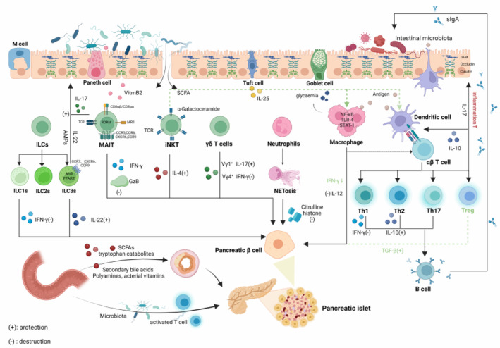
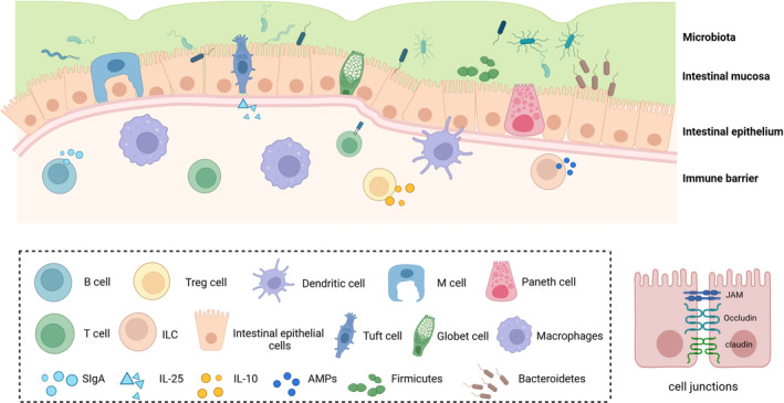
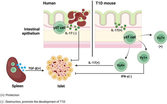
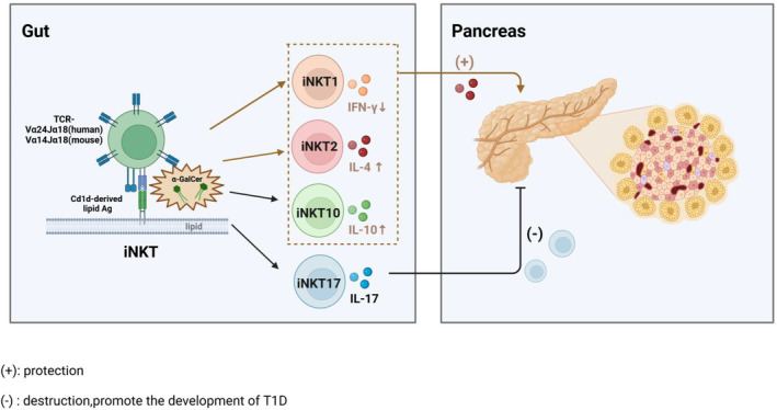
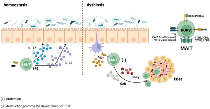
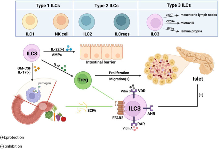
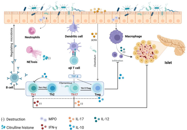
Type 1 diabetes (T1D) is a chronic autoimmune disease characterized by T cell-mediated pancreatic β cell loss, resulting in lifelong absolute insulin deficiency and hyperglycaemia. Environmental factors are recognized as a key contributor to the development of T1D, with the gut serving as a primary interface for environmental stimuli. Recent studies have revealed that the alterations in the intestinal microenvironment profoundly affect host immune responses, contributing to the aetiology and pathogenesis of T1D. However, the dominant intestinal immune cells and the underlying mechanisms remain incompletely elucidated. In this review, we provide an overview of the possible mechanisms of the intestinal mucosal system that underpin the pathogenesis of T1D, shedding light on the roles of both non-classical and classical immune cells in T1D. Our goal is to gain insights into how modulating these immune components may hold potential implications for T1D prevention and provide novel perspectives for immune-mediated therapy.
Keywords: gut‐pancreas axis; intestinal mucosal immunity; microbiota; type 1 diabetes; unconventional T cell.
© 2024 The Author(s). Diabetes, Obesity and Metabolism published by John Wiley & Sons Ltd.
Conflict of interest statement
The authors declare that there is no conflict of interest.
Figures
Similar articles
-
Immune recognition and response to the intestinal microbiome in type 1 diabetes.J Autoimmun. 2016 Jul;71:10-8. doi: 10.1016/j.jaut.2016.02.004. Epub 2016 Feb 20. J Autoimmun. 2016. PMID: 26908163 Review.
-
Cytotoxic and regulatory roles of mucosal-associated invariant T cells in type 1 diabetes.Nat Immunol. 2017 Dec;18(12):1321-1331. doi: 10.1038/ni.3854. Epub 2017 Oct 9. Nat Immunol. 2017. PMID: 28991267 Free PMC article.
-
Gut mucosa alterations and loss of segmented filamentous bacteria in type 1 diabetes are associated with inflammation rather than hyperglycaemia.Gut. 2022 Feb;71(2):296-308. doi: 10.1136/gutjnl-2020-323664. Epub 2021 Feb 16. Gut. 2022. PMID: 33593807
-
On the pathogenesis of insulin-dependent diabetes mellitus: the role of microbiota.Immunol Res. 2017 Feb;65(1):242-256. doi: 10.1007/s12026-016-8832-8. Immunol Res. 2017. PMID: 27421719 Review.
-
Modulation of Gut Microbiota by Low Methoxyl Pectin Attenuates Type 1 Diabetes in Non-obese Diabetic Mice.Front Immunol. 2019 Jul 30;10:1733. doi: 10.3389/fimmu.2019.01733. eCollection 2019. Front Immunol. 2019. PMID: 31417546 Free PMC article.
Cited by
-
Glycemic control and vaccine response: the role of mucosal immunity after vaccination in diabetic patients.Front Immunol. 2025 May 8;16:1577523. doi: 10.3389/fimmu.2025.1577523. eCollection 2025. Front Immunol. 2025. PMID: 40406123 Free PMC article. Review.
-
Gut-tropic α4β7+CD8+ T cells contribute to pancreatic β cell destruction in type 1 diabetes.Front Immunol. 2025 Jul 10;16:1623428. doi: 10.3389/fimmu.2025.1623428. eCollection 2025. Front Immunol. 2025. PMID: 40709181 Free PMC article.
References
-
- Hermann R, Knip M, Veijola R, et al. Temporal changes in the frequencies of HLA genotypes in patients with type 1 diabetes—indication of an increased environmental pressure? Diabetologia. 2003;46(3):420‐425. - PubMed
-
- Söderström U, Aman J, Hjern A. Being born in Sweden increases the risk for type 1 diabetes – a study of migration of children to Sweden as a natural experiment. Acta Paediatr. 2012;101(1):73‐77. - PubMed
-
- Nisticò L, Iafusco D, Galderisi A, et al. Emerging effects of early environmental factors over genetic background for type 1 diabetes susceptibility: evidence from a Nationwide Italian Twin Study. J Clin Endocrinol Metab. 2012;97(8):E1483‐E1491. - PubMed
Publication types
MeSH terms
Grants and funding
LinkOut - more resources
Full Text Sources
Medical
