

Force generation and resistance in human mitosis
- PMID: 39618797
- PMCID: PMC11604895
- DOI: 10.1007/s12551-024-01235-0
Force generation and resistance in human mitosis
Abstract
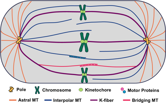
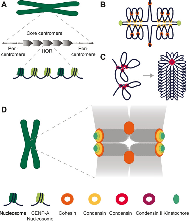
Since the first observations of chromosome segregation over 150 years ago, efforts to observe the forces that drive mitosis have evolved alongside advances in microscopy. The mitotic spindle acts as the major generator of force through the highly regulated polymerization and depolymerization of microtubules as well as associated motor proteins. Centromeric chromatin, along with associated proteins including cohesin and condensin, is organized to resist these forces and ensure accurate chromosome segregation. Microtubules and centromeric chromatin join at the kinetochore, a complex protein superstructure. Ongoing research into the forces generated at the kinetochore-microtubule interface has resulted in a range of estimates for forces necessary to separate chromosomes, from tens to hundreds of piconewtons. Still, the exact magnitude and regulation of these forces remain areas of continuing investigation. Determining the precise forces involved in chromosome segregation is hindered by limitations of current measurement techniques, but advances such as optical tweezers combined with fluorescence microscopy are promising for future research.
Keywords: Centromere; Chromosome; Mitosis; Spindle.
© The Author(s) 2024.
Conflict of interest statement
Competing interestsThe authors declare no competing interests.
Figures

References
-
- Akhmanova A, Kapitein LC (2022) Mechanisms of microtubule organization in differentiated animal cells. Nat Rev Mol Cell Biol 23:541–558. 10.1038/s41580-022-00473-y - PubMed
-
- Allen C, Borisy GG (1974) Structural polarity and directional growth of microtubules of Chlamydomonas flagella. J Mol Biol 90:381–402. 10.1016/0022-2836(74)90381-7 - PubMed
Publication types
LinkOut - more resources
Full Text Sources
