. 2024 Nov 25:17:9683-9696.
doi: 10.2147/JIR.S480528. eCollection 2024.

FSCN1 is a Potential Therapeutic Target for Atherosclerosis Revealed by Single-Cell and Bulk RNA Sequencing

Affiliations

FSCN1 is a Potential Therapeutic Target for Atherosclerosis Revealed by Single-Cell and Bulk RNA Sequencing

Lili Zhang et al. J Inflamm Res. .

Abstract

Background: Atherosclerosis (AS) is the major cause of cardiovascular disease. Using integrated single-cell and bulk RNA sequencing data of atherosclerosis, we aimed to investigate the cell phenotype, intercellular communication, and potential therapeutic target in AS.

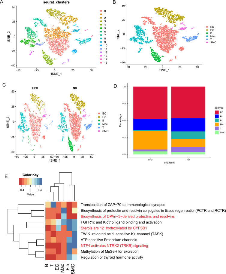
Methods: Single-cell sequencing data from aortic arch of Apoeko mice in normal diet (ND) and high fat diet (HFD) groups (obtained from GSE206239) were analyzed by Seurat, singleR, ReactomeGSA, and cellchat package. scRNA-seq dataset GSE159677 from the carotid artery of the patients with carotid endarterectomy were used to validate the distribution of fascin actin-bundling protein 1 (FSCN1) in cell populations. Bulk RNA sequencing data (GSE43292 and GSE28829) were used to analyzed the expression of FSCN1in AS. A cross-sectional clinical study was utilized to examine the association between FSCN1 and AS. Circulating concentrations of FSCN1 were measured using ELISA kits and assessed using logistic regression analysis and receiver operating characteristic (ROC) curves. Apoeko mice fed with HFD and MAECs treated with oxidized low-density lipoprotein (ox-LDL) were established to detect the expression of FSCN1. Furthermore, we knocked down FSCN1 in MAECs to observe its influence on pyroptosis and migration.

Results: The HFD group had a significantly lower percentage of T cells, fibroblasts, and B cells and a significantly higher percentage of monocytes/macrophages cells. Strong interactions between endothelial cell (EC) and fibroblast in ND groups, while EC interactions with smooth muscle cells (SMC) and T cells were stronger in HFD groups. Semaphorin 7 (SEMA7) mediated signaling pathways were enriched in HFD groups and targeted EC driving by SMC. FSCN1was mainly expressed in EC and had a high expression in human AS samples. The cross-sectional study identified that high level of FSCN1 was associated with increased risk of AS. We also observed that high expression of FSCN1 in ox-LDL-induced MAECs and Apoeko mice fed with HFD. Knockdown of FSCN1 reduced pyroptosis and increased the migration in MAECs.

Conclusion: Knockdown of FSCN1 in EC could alleviate the development and progression of AS. FSCN1 may be a potential prognostic biomarker and therapeutic target in AS.

Keywords: FSCN1; atherosclerosis; endothelial cells; migration; pyroptosis.


Conflict of interest statement

The authors have no relevant financial or non-financial interests to disclose.

Figures

Figure 1
Figure 1
Flow chart of the present study.
Figure 2
Figure 2
Overview of cell populations was profiled. (A) t-distributed stochastic neighbor embedding (t-SNE) represents the aligned gene expression data in ND and HFD groups. (B and C) t-SNE shows the identified major aortic cell types in ND and HFD groups. Monocytes/macrophages: Mac; smooth muscle cells: SMC; endothelial cell: EC; T cells: T; B cells: B; fibroblasts: Fib. (D) The proportions of various cell types in ND and HFD groups. (E) Functional enrichment analysis for the identified hub cell types using “ReactomeGSA” package.
Figure 3
Figure 3
Contiune.
Figure 3
Figure 3
Global cell–cell communication signals participated in atherosclerosis. (A and B) The line width represents the interaction number and interaction weight among hub cell types in ND and HFD groups. (C) Significant signaling of hub cell types in outgoing communication patterns in ND and HFD groups. (D) Bar plot illustrating significant signaling pathways in ND and HFD groups. (E) Circle plot depicting sources and targets for SEMA7 signaling. (F and G) Summary dot plot of ligand-receptor interactions.
Figure 4
Figure 4
Expression of FSCN1 in atherosclerosis. (A) Gene expression patterns projected onto t-SNE plots of FSCN1 (scale: log-transformed gene expression). (B) Gene expression patterns projected onto Violin plots of FSCN1 (scale: log-transformed gene expression). (C) t-SNE plot of aggregate cells from the carotid artery of the patients with carotid endarterectomy with colors denoting different cell clusters in scRNA-seq data (GSE159677). (D) Gene expression patterns projected onto t-SNE plots of FSCN1 (scale: log-transformed gene expression). (E) Gene expression patterns projected onto Violin plots of FSCN1 (scale: log-transformed gene expression). (F) Relative FSCN1 expression in atheroma plaque vs Macroscopically intact tissue in BulkRNA-seq data (GSE43292). (G) Relative FSCN1 expression in advanced atherosclerotic plaque vs early atherosclerotic plaque in RNA-seq data (GSE28829). (H) FSCN1-binding proteins obtained by the STRING tool. (I) ELISA results showing concentrations of FSCN1 in serum from CHD and NO-CHD patients. ****P<0.0001, compared with the NO-CHD group.
Figure 5
Figure 5
The expression of FSCN1 during AS in vivo and in vitro. (A) The representative images of Oil red O-staining of en face aorta. **P<0.01, compared with Apoeko group. (B) Representative images of aortic root sections stained with H&E staining and FSCN1 expression in aortic macrophages of Apoeko hyperlipidemic and ApoeWT mice by co-staining with ECs marker CD31. Scale bars are 200 μm (H&E staining) and 50μm (Immunofluorescence). (C) quantitative data for FSCN1+CD31+cells. ***P<0.001, compared with Apoeko group. (D and E) MAECs treated with ox‑LDL or PBS for 24 h. The protein levels were determined by western analyses. *P<0.05, compared with the PBS group.
Figure 6
Figure 6
Effect of FSCN1 on MAECs pyroptosis and migration. (A) Western blot was used to detect the knockdown efficiency of FSCN1 with three unique siRNAs. (B) The relative expression of the target protein was measured by Image J software. **P<0.01, compared with the siNC group. (C) The pyroptosis-related protein expression levels were measured by Western blotting analysis. (D) NLRP3; (E) GSDMD; (F) caspase‑1; (G) IL‑1β. β-actin levels were used as an internal control. The histogram reports mean ± SEM of protein band density from three experiments (normalized by comparison with β-actin). (H and I) Scratch wound assay was used to evaluate the cell migration capacity. (J and K) Representative images of transwell migration assay. *P<0.05, **P<0.01, ***P<0.001, ****P<0.0001, compared with the siNC group.

Similar articles

References

    1. Virani SS, Alonso A, Aparicio HJ. et al. Heart Disease and Stroke Statistics-2021 Update: a Report From the American Heart Association. Circulation. 2021;143(8):e254–e743. doi:10.1161/CIR.0000000000000950 - DOI - PubMed
    1. Chen W, Li L, Wang J, et al. The ABCA1-efferocytosis axis: a new strategy to protect against atherosclerosis. Clin Chim Acta. 2021;518:1–8. doi:10.1016/j.cca.2021.02.025 - DOI - PubMed
    1. Bonacina F, Di Costanzo A, Genkel V, et al. The heterogeneous cellular landscape of atherosclerosis: implications for future research and therapies. A collaborative review from the EAS young fellows. Atherosclerosis. 2023;372:48–56. doi:10.1016/j.atherosclerosis - DOI - PubMed
    1. Zhang L, Wu X, Hong L. Endothelial Reprogramming in Atherosclerosis. Bioengineering. 2024;11(4):325. doi:10.3390/bioengineering11040325 - DOI - PMC - PubMed
    1. Mause SF, Ritzel E, Deck A, Vogt F, Liehn EA. Engagement of the CXCL12-CXCR4 Axis in the Interaction of Endothelial Progenitor Cell and Smooth Muscle Cell to Promote Phenotype Control and Guard Vascular Homeostasis. Int J Mol Sci. 2022;23(2):867. doi:10.3390/ijms23020867 - DOI - PMC - PubMed