

Cardiac Magnetic Resonance Imaging Findings in COVID-19: Experience from a Tertiary Care Center of North India
- PMID: 39619102
- PMCID: PMC11604198
- DOI: 10.4103/heartviews.heartviews_123_23
Cardiac Magnetic Resonance Imaging Findings in COVID-19: Experience from a Tertiary Care Center of North India
Abstract
Purpose: Here, we describe cardiac magnetic resonance imaging (CMR) findings in patients with proven COVID-19 infection and presenting with cardiac problems both at presentation and in convalescence from a tertiary care center, in North India. A pertinent review of the literature is also discussed.
Materials and methods: Retrospective analysis of patients with real-time reverse transcriptase-polymerase chain reaction proven COVID-19 infection either at presentation or convalescence referred for CMR at our facility from January 2021 to December 2023 was done. CMR was performed on a 3T system (Ingenia, Philips Healthcare, Best, The Netherlands) and examinations were customized according to the clinical indications.
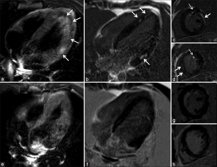
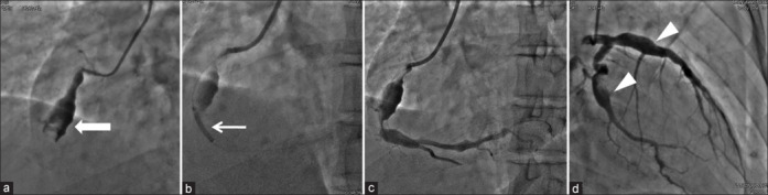
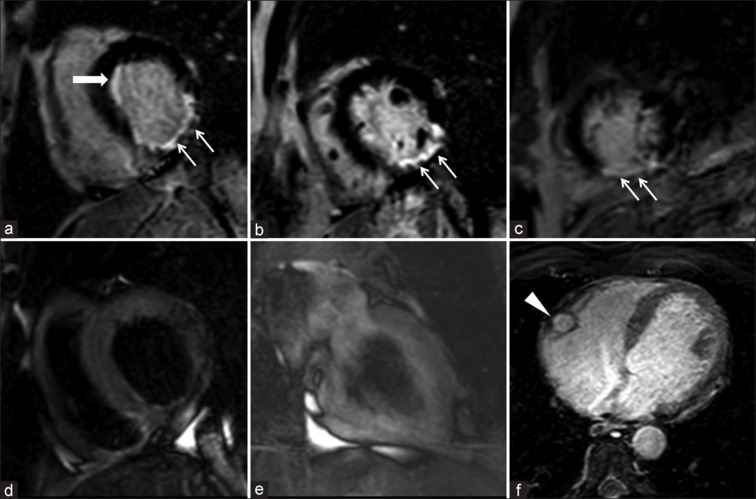
Results: Retrospective analysis yielded 14 patients (4 at presentation; 10 in convalescence). Patients at presentation 4/14 had clinically presented with chest pain with raised troponins and electrographic abnormalities, while 10/14 patients had presented with clinical features of heart failure with two-dimensional transthoracic echocardiography demonstrating systolic dysfunction with reduced left ventricular ejection fraction. Out of 14, 4 patients at presentation, CMR showed features of acute myocarditis in three patients, while one had inferior wall myocardial infarction (MI) (this patient on catheter angiogram had aneurysmally dilated coronary arteries with thrombus and stenosis in the mid right coronary artery which was successfully stented). Out of 14, 10 patients on CMR had features of dilated cardiomyopathy (DCMP).
Conclusion: Cardiac involvement in COVID-19 can have vivid clinicoradiological presentations with features of myocarditis and MI at presentation or DCMP in convalescence. CMR in such cases is a problem-solving tool where myocarditis is candidly differentiated from MI. Moreover, follow-up CMR demonstrates temporal changes in COVID-19-associated myocarditis and evaluation of cardiac structure and function in patients presenting with DCMP.
Keywords: Acute coronary syndrome; cardiac magnetic resonance imaging; cardiovascular complications of COVID-19; coronary artery aneurysm; myocardial infarction; myocarditis.
Copyright: © 2024 Heart Views.
Conflict of interest statement
There are no conflicts of interest.
Figures
Similar articles
-
Multimodality imaging evaluation of Chagas disease: an expert consensus of Brazilian Cardiovascular Imaging Department (DIC) and the European Association of Cardiovascular Imaging (EACVI).Eur Heart J Cardiovasc Imaging. 2018 Apr 1;19(4):459-460n. doi: 10.1093/ehjci/jex154. Eur Heart J Cardiovasc Imaging. 2018. PMID: 29029074
-
Early cardiac magnetic resonance imaging in troponin-positive acute chest pain and non-obstructed coronary arteries.Heart. 2020 Jul;106(13):992-1000. doi: 10.1136/heartjnl-2019-316295. Epub 2020 May 23. Heart. 2020. PMID: 32447308 Free PMC article.
-
Myocarditis as a Possible Underlying Cause for Mid-Ventricular Takotsubo Cardiomyopathy: A Case Report.Cureus. 2024 Dec 16;16(12):e75813. doi: 10.7759/cureus.75813. eCollection 2024 Dec. Cureus. 2024. PMID: 39822402 Free PMC article.
-
Coronary computed tomography angiography (CCTA) and cardiac magnetic resonance (CMR) imaging in the assessment of patients presenting with chest pain suspected for acute coronary syndrome.Ann Transl Med. 2016 Jul;4(13):255. doi: 10.21037/atm.2016.06.30. Ann Transl Med. 2016. PMID: 27500156 Free PMC article. Review.
-
Detection of cardiovascular disease in elite athletes using cardiac magnetic resonance imaging.Rofo. 2013 Dec;185(12):1167-74. doi: 10.1055/s-0033-1350130. Epub 2013 Jul 29. Rofo. 2013. PMID: 23897528 Review.
References
-
- Bonow RO, Fonarow GC, O’Gara PT, Yancy CW. Association of coronavirus disease 2019 (COVID-19) with myocardial injury and mortality. JAMA Cardiol. 2020;5:751–3. - PubMed
-
- Basu-Ray I, Almaddah NK, Adeboye A, Vaqar S, Soos MP. StatPearls. Treasure Island (FL): StatPearls Publishing; 2024. Cardiac Manifestations of Coronavirus (COVID-19) - PubMed
LinkOut - more resources
Full Text Sources