. 2024 Nov 15:15:1483637.
doi: 10.3389/fmicb.2024.1483637. eCollection 2024.

Mycosynthesis of silver nanoparticles from endophytic Aspergillus parasiticus and their antibacterial activity against methicillin-resistant Staphylococcus aureus in vitro and in vivo

Affiliations

Mycosynthesis of silver nanoparticles from endophytic Aspergillus parasiticus and their antibacterial activity against methicillin-resistant Staphylococcus aureus in vitro and in vivo

Enas M Ali et al. Front Microbiol. .

Abstract

Background: Methicillin-resistant Staphylococcus aureus (MRSA) is a drug-resistant and biofilm-forming pathogenic bacteria with severe morbidity and mortality. MRSA showed resistance against currently available antibiotics. Thus, there is an urgent need to develop novel effective treatments with minimal side effects to eliminate MRSA.

Aim: In this study, we aimed to mycosynthesize silver nanoparticles (AgNPs) using the endophytic fungus Aspergillus parasiticus isolated from leaves of Reseda Arabica and to examine their antibacterial activity against MRSA.

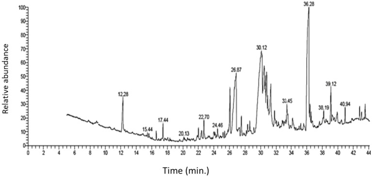
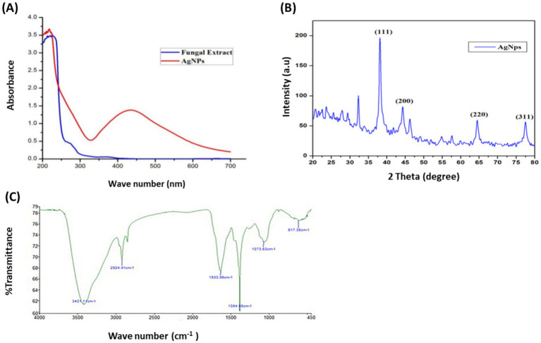
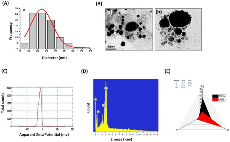
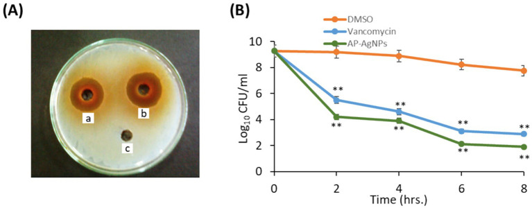
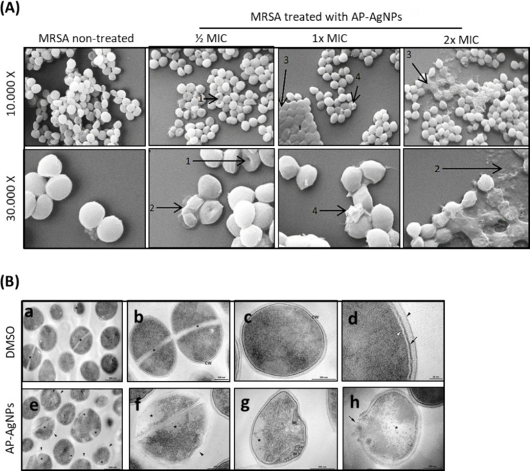
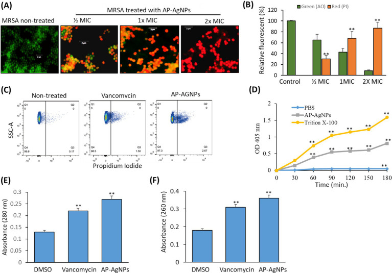
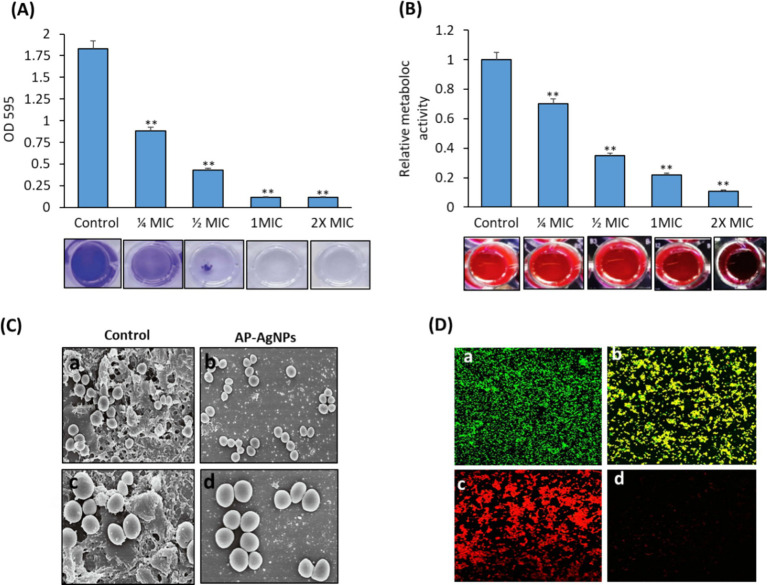
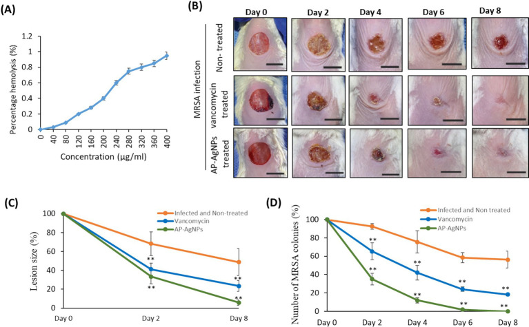
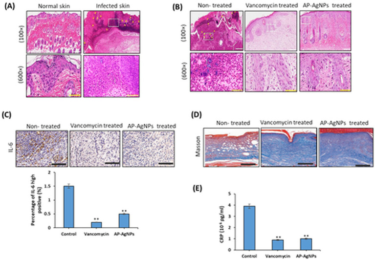
Results: Screening of fungal secondary metabolites using gas chromatography-mass spectroscopy (GC-MS) analysis revealed the presence of high content of bioactive compounds with antibacterial activities. AP-AgNPs were mycosynthesized for the first time using ethyl acetate extract of A. parasiticus and characterized by imaging (transmission electron microscopy (TEM), UV-Vis spectroscopy, zeta potential, X-ray diffraction (XRD), energy-dispersive X-ray analysis (EDX), and Fourier transform infrared spectroscopy (FTIR)). The agar well diffusion method revealed the antibacterial activity of AP-AgNPs against MRSA with 25 μg/mL of minimum inhibitory concentration (MIC). AP-AgNPs were shown to exert antibacterial action via a bactericidal mechanism based on flow cytometry, scanning electron microscopy, and transmission electron microscopy assessment. Our data demonstrated the effective interaction of AP-AgNPs with the bacterial cell membrane, which resulted in cell membrane damage and disruption of cell surface structure. Furthermore, AP-AgNPs successfully prevented the development of MRSA biofilms by disturbing cell adhesion and destructing mature biofilm reaching over 80% clearance rate. Interestingly, topical application of AP-AgNPs to superficial skin infection induced by MRSA in mice effectively promoted wound healing and suppressed bacterial burden.

Conclusion: Our results provide a novel green nanoparticle drug design with effective therapeutic potential against MRSA-induced skin infection.

Keywords: Aspergillus parasiticus; MRSA; silver nanoparticles; skin infection; wound healing.


Conflict of interest statement

The authors declare that the research was conducted in the absence of any commercial or financial relationships that could be construed as a potential conflict of interest. The author(s) declared that they were an editorial board member of Frontiers, at the time of submission. This had no impact on the peer review process and the final decision.

Figures

Figure 1
Figure 1
GC–MS chromatogram of ethyl acetate extract of A. parasiticus.
Figure 2
Figure 2
Confirmation of biosynthesized silver nanoparticles (AP-AgNPs). (A) UV–Vis spectrum of fungal filtrate and AP-AgNPs that were synthesized by the A. parasiticus. The peak values are for the UV–Vis plotted between AP-AgNPs and absorbance ratios. The highest absorbance peak of AP-AgNPs was at approximately 448 nm, corresponding to the Plasmon resonance of AP-AgNPs. (B) XRD spectrum recorded for AP-AgNPs showed four distinct diffraction peaks at 38.19°, 44.37°, 64.56°, and 77.47° indexed 2-theta (degree) values of (111), (200), (220), and (311) crystalline planes of cubic Ag. (C) FTIR spectrum of silver nanoparticles synthesized by A. parasiticus showed peaks at 3,421.1, 2,924.41, 1,633.96, 1,384.56, 1,073.63, and 617.36 cm−1. Those peaks were, respectively, attributed to N-H stretching of primary amine of the protein, alkane C-H stretching, the stretching of conjugated alkane C=C, methylene tails of the protein (CH3-R), C-N of aliphatic amines of polyphenols, and O-H stretching.
Figure 3
Figure 3
Characterization of AP-AgNPs. (A) Histogram of the particle diameter size distribution of the AP-AgNPs based on TEM image analysis. Red line: Gaussian distribution fit. (B) [a] Transmission electron microscopy (TEM) images of AP-AgNPs (scale bar = 100 nm). [b] High-resolution TEM image of nanoparticle of AP-AgNPs (scale bar = 20 nm). According to TEM, AP-AgNPs are mostly spherical, and the average particle size is 27.7 nm. The diameter range is 5.77–73.14 nm. The geometric mean equal value of 24.96 ± 1.65 nm. (C) Zeta potential of AP-AgNPs. The zeta potential value was −25.5 ± 3.15 mv. The negative value confirmed the stability of AP-AgNPs. (D) EDX images and (E) weight percentage of elements. The spectrum displayed that silver, constituting 53.29% of the total, was the greatest important element, followed by carbon (33.5%) and oxygen (13.21%), representing the establishment of AP-AgNPs.
Figure 4
Figure 4
Antibacterial potential of AP-AgNPs. (A) Agar well diffusion method showing the antibacterial activity depicting the zone of antibacterial inhibition against MRSA by vancomycin (positive control) (50 μg/mL) (a), AP-AgNPs (25 μg/mL) (b), and negative control DMSO (c). Data are expressed as the mean zone of inhibition in millimeters. (B) Time–kill curves of MRSA following exposure to AP-AgNPs, vancomycin, and DMSO. Non-treated MRSA cells were used as a control. There was a decrease in the rate of cell growth when MRSA cells were treated with AP-AgNPs compared to the control cells. Values are means ± SD (**p < 0.005, compared to control DMSO-treated cells).
Figure 5
Figure 5
Effect of AP-AgNPs on MRSA ultrastructure. (A) Scanning electron microscopy of MRSA cells after 24 h of exposure to AP-AgNPs in nutrient broth. The cleavage sites (1), cellular debris (2), boss-like protuberances (3), and unusual protrusions (4) are shown by arrows. (B) Transmission electron microscopy of MRSA. (a–d) showing cells treated with DMSO (control). (a) General view displays rounded cells with a thick cell wall envelope and homogeneous electron density in the cytoplasm. Septa (*) appears in some cells. (b,c) Detailed view of non-dividing (b) and dividing (c) cells. A tripartite cell wall (CW) is seen enclosing the plasma membrane. (d) Inset of the cell wall. The black arrowhead specifies the outer highly stained fibrous surface and intermediate translucent region; arrow points to a heavily stained inner thin zone; the plasma membrane (white arrowhead) is seen below this electron-dense layer of the wall. (e–f) Showing cells treated with AP-AgNPs (MIC = 25 μg/mL) (M/4). (e) General view: some cells showed abnormal electron density in the cytoplasm and altered cell wall with no visible tripartite layers (black arrowheads). (f) Inset of figure e. (g) Detailed view of a cell showing alterations in the shape, loss of cytosolic electron density, and mesosome-like structures (M). (h) A lysed cell with cell wall disruption (arrow) and cytoplasmic disintegration (*).
Figure 6
Figure 6
Effect of AP-AgNPs on MRSA cell membrane. (A) Fluorescent confocal microscopy of live and dead bacteria MRSA after 24 h of exposure to AP-AgNPs in nutrient broth and stained with propidium iodide (red color for dead bacteria) and acridine orange (green color for live bacteria). (B) Relative fluorescence intensity of PI and AO at different treatments. (C) Analysis of cell membrane integrity of MRSA by FACS analysis using PI staining under different treatments. (D) Effects of AP-AgNPs on the permeability of the cell membrane of MRSA. The membrane permeability was determined by the optical density value. AP-AgNPs and Triton X-100 increased the membrane permeability of MRSA, as compared with PBS (p < 0.01). (E) Leakage of intracellular proteins. (F) Leakage of intracellular nucleic acids. Values are means ± SD (*p < 0.05, **p < 0.005, compared to DMSO or PBS-treated control).
Figure 7
Figure 7
Effect of AP-AgNPs on biofilm formation by MRSA. (A) Crystal violet and (B) resazurin (excitation wavelength: 570 nm; emission wavelength: 600 nm). Values are means ± SD (**p < 0.005, compared to control cells). (C) Scanning electron microscopy images of biofilm formation of MRSA treated with (a,c) DMSO (control) or (b,d) AP-AgNPs at 35°C for 24 h. A significant bacterial biofilm growth, which led to clusters with composite morphology, was seen on the surface of the glass in the control group. However, limited bacterial biofilm formation was seen on the surface of the (AP-AgNPs) treatment group. (D) Confocal laser scanning microscopy image of LIVE/DEADH-stained demonstrating the effects of different AP-AgNP concentrations on MRSA biofilm formation. Biofilms were formed on cover slides within 48 h at 37°C. Biofilms were treated with AP-AgNPs for 48 h at 37°C. (a) Control (DMSO); (b–d) treatment with AP-AgNPs at 25 μg/mL, 50 μg/mL, or 100 μg/mL, respectively. Green, viable cells; Red, dead cells.
Figure 8
Figure 8
Mouse superficial wound infection. (A) Hemolytic activity of AP-AgNPs. The hemolytic activity of AP-AgNPs was estimated by monitoring the increase in the absorbance at 570 nm after incubating human red blood cells with different AP-AgNP concentrations at 37°C for 1 h. The positive control was 0.1% Triton X-100, and DMSO was used as a blank. (B) Mouse superficial wounds were performed by repeated tape stripping to remove the epidermis. Wounds were infected with 1 × 107 MRSA colonies and treated every day with 10% ethanol in propylene glycol (non-treated control), 25 μg/mL AP-AgNPs (MIC) in 10% ethanol in propylene glycol or 50 μg/mL vancomycin (MIC) for eight days. The wounds of all mice were snapped on days 0–8. Scale bar: 1 cm. (C) Lesion size analysis on days 2 and 8 after initial surgery. (D) Numbers of MRSA colonies isolated from infected superficial mouse wounds under different treatments. Data are expressed as means ± SD (n = 6 mice/group) (**p < 0.005, compared to control infected and non-treated mice).
Figure 9
Figure 9
Histology of wounds with H&E staining. Mice were sacrificed and tissues were cut into 6-μm sections, stained with H&E, and mounted. (A) Skin sections comparing normal skin versus MRSA-infected skin after 2 days of infection. (B) MRSA-infected skin sections from mice non-treated or treated with vancomycin or AP-AgNPs after 8 days of treatment. Magnification = 100X and 600X. (C) Immunohistochemical staining for IL-6 in the hypodermis from the infected wounds on day 8; scale bar: 100 μm. (D) Masson’s staining of the tissues from the infected wounds on day 8; scale bar: 200 μm. (E) serum C-reactive protein (CRP) levels. Data are expressed as means ± SD (n = 6 mice/group) (**p < 0.005, compared to control infected and non-treated mice).

Similar articles

Cited by

References

    1. Abada E., Mashraqi A., Modafer Y., Al Abboud M. A., El-Shabasy A. (2024). Review green synthesis of silver nanoparticles by using plant extracts and their antimicrobial activity. Saudi J Biol Sci 31:103877. doi: 10.1016/j.sjbs.2023.103877, PMID: - DOI - PMC - PubMed
    1. Abtin A., Jain R., Mitchell A. J., Roediger B., Brzoska A. J., Tikoo S., et al. . (2014). Perivascular macrophages mediate neutrophil recruitment during bacterial skin infection. Nat. Immunol. 15, 45–53. doi: 10.1038/ni.2769, PMID: - DOI - PMC - PubMed
    1. Agoramoorthy G., Chandrasekaran M., Venkatesalu V., Hsu M. J. (2007). Antibacterial and antifungal activities of fatty acid methyl esters of the blind-your-eye mangrove from India. Braz. J. Microbiol. 38, 739–742. doi: 10.1590/S1517-83822007000400028 - DOI
    1. Agrawal S., Nandeibam J., Sarangthem I. (2021). Ultrastructural changes in methicillin-resistant Staphylococcus aureus (MRSA) induced by metabolites of thermophilous fungi Acrophialophora levis. PLoS One 16:e0258607. doi: 10.1371/journal.pone.0258607, PMID: - DOI - PMC - PubMed
    1. Ahamad I., Bano F., Anwer R., Srivastava P., Kumar R., Fatma T. (2021). Antibiofilm activities of biogenic silver nanoparticles against Candida albicans. Front. Microbiol. 12:741493. doi: 10.3389/fmicb.2021.741493, PMID: - DOI - PMC - PubMed

LinkOut - more resources