

Species Identification and Genotyping of Cutaneous Leishmaniasis in Clinical Samples Based on ITS1-PCR-Sequencing in Southeast Iran
- PMID: 39619911
- PMCID: PMC11607147
- DOI: 10.18502/ijph.v53i11.16962
Species Identification and Genotyping of Cutaneous Leishmaniasis in Clinical Samples Based on ITS1-PCR-Sequencing in Southeast Iran
Abstract
Background: Cutaneous leishmaniasis (CL) is one of the most common parasitic diseases in many regions of Iran. It has a major role in deprived societies. We aimed to identify Leishmania species based on molecular as ITS1-rDNA-PCR internal transcribed spacer 1 (ITS1) region, microscopy, and culture techniques in diagnosing cutaneous leishmaniasis.
Methods: From April 2018 to May 2020, we conducted a cross-sectional study involving 32 patients with suspected CL lesions in Sistan and Baluchistan Province, located in southeast Iran. Samples were subjected to microscopic examination, culture, and PCR amplification targeting the internal transcribed spacer 1 (ITS1) region. DNA sequencing was performed on PCR-positive samples for species identification and phylogenetic analysis.
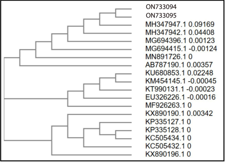
Results: PCR demonstrated superior sensitivity (93.75%, 30/32) compared to culture (56.25%, 18/32) and microscopic examination (53.1%, 17/32). Molecular analysis revealed that L. major was the predominant causative agent of CL in the study area, with L. tropica occurring less frequently. Sequencing and phylogenetic analysis of the ITS1 region showed high intraspecies similarity among L. tropica isolates, while L. major isolates exhibited greater genetic diversity.
Conclusion: This study shows the co-existence of L. major and L. tropica in Mirjaveh, southeast Iran, with L. major as the primary cause. While L. tropica isolates displayed high genetic similarity, L. major samples were more diverse, indicating different epidemiological patterns. These findings highlight the importance of molecular methods for accurately identifying Leishmania species and understanding CL epidemiology in the region.
Keywords: Cutaneous leishmaniasis; Iran; Polymerase chain reaction; Sequence.
Copyright© 2024 Dabirzadeh et al. Published by Tehran University of Medical Sciences.
Figures


Similar articles
-
Genetic diversity of Leishmania tropica strains isolated from clinical forms of cutaneous leishmaniasis in rural districts of Herat province, Western Afghanistan, based on ITS1-rDNA.Infect Genet Evol. 2016 Jul;41:120-127. doi: 10.1016/j.meegid.2016.03.031. Epub 2016 Apr 5. Infect Genet Evol. 2016. PMID: 27063410
-
Molecular Diagnosis of Clinical Isolates of Cutaneous Leishmaniasis Using ITS1 and KDNA Genes and Genetic Polymorphism of Leishmania in Kashan, Iran.Pak J Biol Sci. 2016;19(3):136-142. doi: 10.3923/pjbs.2016.136.142. Pak J Biol Sci. 2016. PMID: 29023050
-
Detection, molecular typing and phylogenetic analysis of Leishmania isolated from cases of leishmaniasis among Syrian refugees in Lebanon.Parasite Epidemiol Control. 2016 Feb 27;1(2):159-168. doi: 10.1016/j.parepi.2016.02.002. eCollection 2016 Jun. Parasite Epidemiol Control. 2016. PMID: 29988171 Free PMC article.
-
Molecular Identification of Agents of Human Cutaneous Leishmaniasis and Canine Visceral Leishmaniasis in Different Areas of Iran Using Internal Transcribed Spacer 1 PCR-RFLP.J Arthropod Borne Dis. 2018 Jun 13;12(2):162-171. eCollection 2018 Jun. J Arthropod Borne Dis. 2018. PMID: 30123810 Free PMC article.
-
The Geographical Distribution of Human Cutaneous and Visceral Leishmania Species Identified by Molecular Methods in Iran: A Systematic Review With Meta-Analysis.Front Public Health. 2021 Jun 25;9:661674. doi: 10.3389/fpubh.2021.661674. eCollection 2021. Front Public Health. 2021. PMID: 34249836 Free PMC article.
References
-
- Gutiérrez V, Seabra AB, Reguera RM, Khandare J, Calderón M. (2016). New approaches from nanomedicine for treating leishmaniasis. Chem Soc Rev, 45(1): 152–168. - PubMed
-
- WHO Expert Committee on the Control of the Leishmaniases & World Health Organization . (2010). Control of the leishmaniases: report of a meeting of the WHO Expert Commitee on the Control of Leishmaniases, Geneva, 22–26 March 2010. World Health Organization.
-
- Alemayehu B, Alemayehu M. (2017). Leishmaniasis: a review on parasite, vector, and reservoir host. Health Sci J, 11(4): 519.
-
- Moreira OC, Yadon ZE, Cupolillo E. (2018). The applicability of real-time PCR in the diagnostic of cutaneous leishmaniasis and parasite quantification for clinical management: current status and perspectives. Acta Trop, 184: 29–37. - PubMed
LinkOut - more resources
Full Text Sources
Miscellaneous