Review
. 2024 Nov 15:12:1455140.
doi: 10.3389/fcell.2024.1455140. eCollection 2024.

Evaluation of mesenchymal stem cells as an in vitro model for inherited retinal diseases

Affiliations
Review

Evaluation of mesenchymal stem cells as an in vitro model for inherited retinal diseases

Maria Dodina et al. Front Cell Dev Biol. .

Abstract

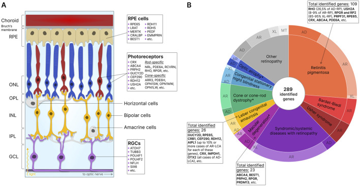
Retinal pathologies are major causes of vision impairment and blindness in humans, and inherited retinal diseases (IRDs), such as retinitis pigmentosa, Leber congenital amaurosis, and Stargardt disease, greatly contribute to this problem. In vitro disease modeling can be used for understanding the development of pathology and for screening therapeutic pharmaceutical compounds. In the preclinical research phase, in vitro models complement in vivo models by reducing animal studies, decreasing costs, and shortening research timelines. Additionally, animal models may not always accurately replicate the human disease phenotype. This review examines the types of cells that can be used to create in vitro IRD models, including retina-specific cell lines, primary retinal cells, induced pluripotent stem cells (iPSCs), and more. Special attention is given to mesenchymal stem cells (MSCs), which are characterized by various isolation sources, relative ease of isolation, and straightforward differentiation. MSCs derived from bone marrow (BM), adipose tissue (AT), dental tissue (DT), umbilical cord (UC), and other sources can differentiate into retinal cells, including photoreceptor cells and retinal pigment epithelial (RPE) cells, dysfunction of which is most commonly associated with IRDs. Subsequent differentiation of MSCs into retinal cells can be carried out via various methods: culturing in induction media supplemented with certain growth factors, co-culturing with retinal cells or in their conditioned media, or regulating gene expression with viral vector-delivered transcription factors (TFs) or microRNAs (miRNAs). Compared to the popular iPSCs, for example, MSC-based models are significantly cheaper and faster to obtain, making them more feasible for large-scale drug screening. Nevertheless, the existing differentiation methods need further optimization for this promising platform to receive the success it deserves.

Keywords: IRD; MSC; in vitro disease modeling; inherited retinal diseases; mesenchymal stem cells; retinal cells.


Conflict of interest statement

The authors declare that the research was conducted in the absence of any commercial or financial relationships that could be construed as a potential conflict of interest.

Figures

FIGURE 1
FIGURE 1
Retinal structure and IRD-associated genes expressed in retinal cells. (A) Structure of the retina showing major cells and layers. RPE - retinal pigment epithelium; RGCs - retinal ganglion cells; ONL - outer nuclear layer; OPL - outer plexiform layer; INL - inner nuclear layer; IPL - inner plexiform layer; GCL - ganglion cell layer. Genes specific to photoreceptors, RPE, and RGCs are indicated. (B) IRD-associated genes by disease category. The data for the diagram was obtained from RetNet (Retinal Information Network, 2024). While different mutations in the same gene may be associated with different diseases, each identified gene is counted only once for the first-reported disease (usually the most common disease). The total number of identified genes and the most commonly disease-associated genes are listed by the disease category. The outer circle of the diagram reflects the number of genes associated with different types of inheritance: AD - autosomal dominant; AR - autosomal recessive; XL - X-linked; MT - mitochondrial. *- diseases for which XL-forms are also identified.
FIGURE 2
FIGURE 2
Cell types used in the development of in vitro IRD models. MSCs - mesenchymal stem cells; PSCs - pluripotent stem cells; ESCs–embryonic stem cells; iPSCs–induced pluripotent stem cells. 2D or 3D models can be obtained based on various cell types. *- applicable when not used in combination with organ-on-a-chip and bioprinting technologies.
FIGURE 3
FIGURE 3
MSCs as a potential source of retinal cells. MSCs can be isolated from various sources based on phenotyping markers. Most often used: BMSCs–bone marrow stem cells, ADSCs–adipose-derived stem cells, UC-MSCs–umbilical cord MSCs, WJ-MSCs–Wharton’s jelly MSCs, DPSCs–dental pulp stem cells; PDLSCs–periodontal ligament stem cells, etc. These cells are capable of self-renewal and differentiation in three directions: osteogenic, adipogenic, and chondrogenic. Under certain conditions, they are able to differentiate into retinal pigment epithelial (RPE) cells, photoreceptors, and retinal ganglion cells (RGCs).
FIGURE 4
FIGURE 4
MSCs for in vitro modeling of healthy retinal physiology, or IRDs, and in cell therapy of degenerative retinal diseases. Functional retinal cells can be obtained from the MSCs of a healthy donor and are suitable for studying healthy physiology. Similar to retinal cells obtained from IRD patient MSCs, gene-edited MSCs from healthy donors can be used to study IRD pathology or for drug testing.
FIGURE 5
FIGURE 5
MSC-based clinical trials for retinal degenerative conditions (valid for June 2024). (A) Clinical trials by study phase. NA - not applicable. (B) Study status. (C) Country of study. The largest number of studies are located in Turkey, the United States, and Indonesia. (D) Clinical trials by the condition at which the therapy is aimed. (E) Clinical studies by MSC source. (F) Route of administration.

Similar articles

Cited by

References

    1. Achberger K., Haderspeck J. C., Kleger A., Liebau S. (2019). Stem cell-based retina models. Adv. Drug Deliv. Rev. 140, 33–50. 10.1016/j.addr.2018.05.005 - DOI - PubMed
    1. Adak S., Magdalene D., Deshmukh S., Das D., Jaganathan B. G. (2021). A review on mesenchymal stem cells for treatment of retinal diseases. Stem Cell Rev. Rep. 17 (4), 1154–1173. 10.1007/s12015-020-10090-x - DOI - PMC - PubMed
    1. Afanasyeva T. A. V., Corral-Serrano J. C., Garanto A., Roepman R., Cheetham M. E., Collin R. W. J. (2021). A look into retinal organoids: methods, analytical techniques, and applications. Cell. Mol. Life Sci. 78 (19-20), 6505–6532. 10.1007/s00018-021-03917-4 - DOI - PMC - PubMed
    1. Afflerbach A. K., Kiri M. D., Detinis T., Maoz B. M. (2020). Mesenchymal stem cells as a promising cell source for integration in novel in vitro models. Biomolecules 10 (9), 1306. 10.3390/biom10091306 - DOI - PMC - PubMed
    1. Afshari A., Shamdani S., Uzan G., Naserian S., Azarpira N. (2020). Different approaches for transformation of mesenchymal stem cells into hepatocyte-like cells. Stem Cell Res. Ther. 11 (1), 54. 10.1186/s13287-020-1555-8 - DOI - PMC - PubMed

LinkOut - more resources