. 2024 Dec 2:152:e150.
doi: 10.1017/S0950268824001420.

Origin and evolution of West Nile virus lineage 1 in Italy

Affiliations

Origin and evolution of West Nile virus lineage 1 in Italy

Andrea Silverj et al. Epidemiol Infect. .

Abstract

West Nile virus (WNV) is a mosquito-borne pathogen that can infect humans, equids, and many bird species, posing a threat to their health. It consists of eight lineages, with Lineage 1 (L1) and Lineage 2 (L2) being the most prevalent and pathogenic. Italy is one of the hardest-hit European nations, with 330 neurological cases and 37 fatalities in humans in the 2021-2022 season, in which the L1 re-emerged after several years of low circulation. We assembled a database comprising all publicly available WNV genomes, along with 31 new Italian strains of WNV L1 sequenced in this study, to trace their evolutionary history using phylodynamics and phylogeography. Our analysis suggests that WNV L1 may have initially entered Italy from Northern Africa around 1985 and indicates a connection between European and Western Mediterranean countries, with two distinct strains circulating within Italy. Furthermore, we identified new genetic mutations that are typical of the Italian strains and that can be tested in future studies to assess their pathogenicity. Our research clarifies the dynamics of WNV L1 in Italy, provides a comprehensive dataset of genome sequences for future reference, and underscores the critical need for continuous and coordinated surveillance efforts between Europe and Africa.

Keywords: Europe; Italy; West Nile virus; lineage 1; mosquito; phylogeography.


Conflict of interest statement

The authors declare that they have no competing interests.

Figures
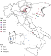
Figure 1.
Figure 1.
Geo-localization of West Nile virus lineage 1 sequenced sample collection sites. The sampling date is indicated by a colour scale. The shape indicates the host from which the sample was extracted. For the map, we used region border shapes from GADM (which are freely available under a CC-BY license; https://gadm.org/license.html), retrieving them through the ‘getData’ function of the ‘raster’ R package (https://www.rdocumentation.org/packages/raster/versions/3.6-20/topics/getData).
Figure 2.
Figure 2.
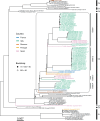
Maximum likelihood tree of West Nile virus lineage 1 sequences. Seven major clusters (C1–C7, bar on the right) can be identified. Mediterranean countries that are part of a major cluster that includes all Italian genomes (all part of the group that was used for the molecular clock and phylogeographic analysis, highlighted in a black square with rounded corners) are indicated by different colours. Inside this group, 4 Italian clades (Clade 1–4) can be defined based on the topology of the tree. Bootstrap support values (BS) >90 are not shown; bootstrap support values between 70 and 90 are represented by a full black circle; low bootstrap supports (BS < 50) are represented with a white circle. Sequences obtained in this study are indicated by the “*” symbol. A scale bar representing substitutions per site is shown at the bottom left part of the figure.
Figure 3.
Figure 3.
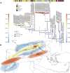
Phylogeographic analysis of the WNV L1 Western Mediterranean clade. (A) Molecular clock of the clade including all Italian sequences. Posterior probabilities of the nodes are indicated by different shapes. The bar at the top indicates the host from which the genome was isolated (human, bird, mosquito, or horse). The time scales on the right and left sides indicate, with a colour gradient, the sampling time (blue: older; red: more recent) of the genomes (see tips) or the median estimated age of their ancestors (see nodes). The four main introductions in Italy are indicated by numbers close to the nodes (1–4), while genomes from the recent outbreaks in Southern Italy (SI) and Northern Italy (NI) are highlighted by a dotted square and a full square, respectively. (B) Phylogeographic diffusion pattern in continuous scale of the virus in the Mediterranean basin. Genome sampling sites and reconstructed locations are plotted together on a map, showing the connections among all genomes and their inferred ancestors. Again, the same colour scale used in (A) indicates the sampling time of the genomes or the median estimated age of their ancestors. The coloured areas in the figure represent the 95% HPDs of the locations and times inferred by the model. For the map, we used region border shapes from GADM (which are freely available under a CC-BY license; https://gadm.org/license.html), retrieving them through the ‘getData’ function of the ‘raster’ R package (https://www.rdocumentation.org/packages/raster/versions/3.6-20/topics/getData).
Figure 4.
Figure 4.
Amino acid sequence analysis. Amino acid sequence comparison between West Nile virus lineage 1 Italian strains and other Western-Mediterranean strains included in Cluster 2, other than the 1999 NC_009942 strain representative for the USA genome sequence group included in Cluster 4, is displayed. C, Capsid protein; prM, pre-membrane protein; M, Membrane protein; NS, non-structural proteins. Common point mutations (51-T, 99-T, 96-I) detected in the two Campanian strains OP850023/2022 and MW627239/2022 are displayed in pink; the point mutation 93-K of the Envelope protein detected in the 2021–2022 Northern-Eastern Italian strains and in the 2015 French strain MT863559 is displayed in light green; the 122H amino acid substitution of the NS2a protein detected in the 2021–2022 Northern-Eastern Italian strains is displayed in yellow; the 249-P point mutation is displayed in light blue.

References

    1. Rizzoli A, et al. (2015) The challenge of West Nile virus in Europe: knowledge gaps and research priorities. Euro Surveillance 20, 21135. - PubMed
    1. Chancey C, et al. (2015) The global ecology and epidemiology of West Nile virus. BioMed Research International 2015, 376230. - PMC - PubMed
    1. Kramer LD, et al. (2007). West Nile virus. The Lancet Neurology 6, 171–181. - PubMed
    1. Smithburn KC, et al. (1940) A neurotropic virus isolated from the blood of a native of Uganda. American Journal of Tropical Medicine 20, 471–472.
    1. Pierson TC and Diamond MS (2020) The continued threat of emerging flaviviruses. Nature Microbiology 5, 796–812. - PMC - PubMed