Cerebral hypoperfusion reduces tau accumulation
- PMID: 39621511
- PMCID: PMC11752094
- DOI: 10.1002/acn3.52247
Cerebral hypoperfusion reduces tau accumulation
Abstract
Objective: Alzheimer's disease (AD) often coexists with cerebrovascular diseases. However, the impact of cerebrovascular diseases such as stroke on AD pathology remains poorly understood.
Methods: This study examines the correlation between cerebrovascular diseases and AD pathology. The research was carried out using clinical and neuropathological data collected from the National Alzheimer's Coordinating Center (NACC) database and an animal model in which bilateral common carotid artery stenosis surgery was performed, following the injection of tau seeds into the brains of wild-type mice.
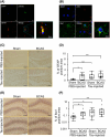
Results: Analysis of the NACC database suggests that clinical stroke history and lacunar infarcts are associated with lower neurofibrillary tangle pathology. An animal model demonstrates that chronic cerebral hypoperfusion reduces tau pathology, which was observed in not only neurons but also astrocytes, microglia, and oligodendrocytes. Furthermore, we found that astrocytes and microglia were activated in response to tau pathology and chronic cerebral hypoperfusion. Additionally, cerebral hypoperfusion increased a lysosomal enzyme, cathepsin D.
Interpretation: These data together indicate that cerebral hypoperfusion reduces tau accumulation likely through an increase in microglial phagocytic activity towards tau and an elevation in degradation through cathepsin D. This study contributes to understanding the relationship between tau pathology and cerebrovascular diseases in older people with multimorbidity.
© 2024 The Author(s). Annals of Clinical and Translational Neurology published by Wiley Periodicals LLC on behalf of American Neurological Association.
Conflict of interest statement
The authors declare no conflicts of interest associated with this manuscript.
Figures


References
MeSH terms
Substances
Grants and funding
- MEXT15K15272/Grants-in-Aid from Japan Promotion of Science; the Japanese Ministry of Education, Culture, Sports, Science, and Technology
- 19-9/Funding for Longevity Sciences from the National Center for Geriatrics and Gerontology
- P20 AG068053/AG/NIA NIH HHS/United States
- P30 AG062421/AG/NIA NIH HHS/United States
- Takeda Medical Research Foundation Research
- P30 AG066508/AG/NIA NIH HHS/United States
- P30 AG072973/AG/NIA NIH HHS/United States
- P30 AG066530/AG/NIA NIH HHS/United States
- Novartis Foundation for Gerontological Research Award
- P30 AG066509/AG/NIA NIH HHS/United States
- P30 AG066546/AG/NIA NIH HHS/United States
- Mitsui Sumitomo Insurance Welfare Foundation
- P30 AG072979/AG/NIA NIH HHS/United States
- P20 AG068082/AG/NIA NIH HHS/United States
- P30 AG072975/AG/NIA NIH HHS/United States
- P30 AG066444/AG/NIA NIH HHS/United States
- P30 AG066507/AG/NIA NIH HHS/United States
- P30 AG072946/AG/NIA NIH HHS/United States
- P30 AG066518/AG/NIA NIH HHS/United States
- P30 AG066511/AG/NIA NIH HHS/United States
- U24 AG072122/AG/NIA NIH HHS/United States
- MEXT21H02844/Grants-in-Aid from Japan Promotion of Science; the Japanese Ministry of Education, Culture, Sports, Science, and Technology
- P30 AG066512/AG/NIA NIH HHS/United States
- MEXT26293167/Grants-in-Aid from Japan Promotion of Science; the Japanese Ministry of Education, Culture, Sports, Science, and Technology
- P30 AG066515/AG/NIA NIH HHS/United States
- P30 AG072978/AG/NIA NIH HHS/United States
- P30 AG062429/AG/NIA NIH HHS/United States
- P30 AG066519/AG/NIA NIH HHS/United States
- Takeda Science Foundation Research Encouragement Grant
- 28-45/Funding for Longevity Sciences from the National Center for Geriatrics and Gerontology
- P30 AG062422/AG/NIA NIH HHS/United States
- R01 AG079280/AG/NIA NIH HHS/United States
- P30 AG066462/AG/NIA NIH HHS/United States
- 19-3/Funding for Longevity Sciences from the National Center for Geriatrics and Gerontology
- 21-12/Funding for Longevity Sciences from the National Center for Geriatrics and Gerontology
- MEXT24K02361/Grants-in-Aid from Japan Promotion of Science; the Japanese Ministry of Education, Culture, Sports, Science, and Technology
- P20 AG068077/AG/NIA NIH HHS/United States
- P30 AG072977/AG/NIA NIH HHS/United States
- P30 AG062677/AG/NIA NIH HHS/United States
- P20 AG068024/AG/NIA NIH HHS/United States
- P30 AG072958/AG/NIA NIH HHS/United States
- P30 AG062715/AG/NIA NIH HHS/United States
- P30 AG066506/AG/NIA NIH HHS/United States
- P30 AG066468/AG/NIA NIH HHS/United States
- Annual Research Award Grant from the Japanese Society of Anti-aging Medicine
- P30 AG072976/AG/NIA NIH HHS/United States
- P30 AG072947/AG/NIA NIH HHS/United States
- P30 AG072931/AG/NIA NIH HHS/United States
- MEXT17H04154/Grants-in-Aid from Japan Promotion of Science; the Japanese Ministry of Education, Culture, Sports, Science, and Technology
- SENSHIN Medical Research Foundation Research Grant
- P30 AG072972/AG/NIA NIH HHS/United States
- P30 AG066514/AG/NIA NIH HHS/United States
- P30 AG072959/AG/NIA NIH HHS/United States
- 24-16/Funding for Longevity Sciences from the National Center for Geriatrics and Gerontology
LinkOut - more resources
Full Text Sources
Medical

