. 2024 Dec 2;19(12):e0300297.
doi: 10.1371/journal.pone.0300297. eCollection 2024.

Discovery of anti-SARS-CoV-2 S2 protein antibody CV804 with broad-spectrum reactivity with various beta coronaviruses and analysis of its pharmacological properties in vitro and in vivo

Affiliations

Discovery of anti-SARS-CoV-2 S2 protein antibody CV804 with broad-spectrum reactivity with various beta coronaviruses and analysis of its pharmacological properties in vitro and in vivo

Yoji Tsugawa et al. PLoS One. .

Abstract

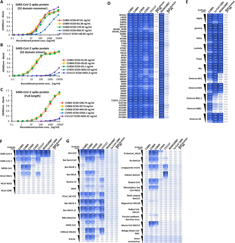
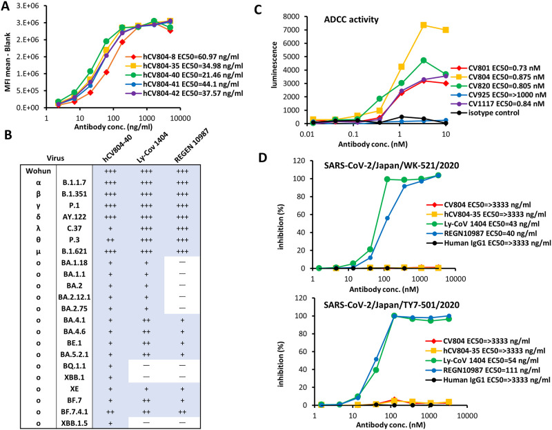
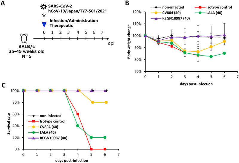
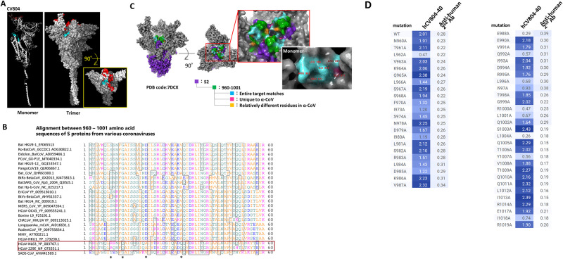
The SARS-CoV-2 pandemic alerted the potential for significant harm due to future cross-species transmission of various animal coronaviruses to human. There is a significant need of antibody-based drugs to treat patients infected with previously unseen coronaviruses. In this study, we generated CV804, an antibody that binds to the S2 domain of SARS-CoV-2 spike protein, which is highly conserved across the coronavirus family and less susceptible to mutations. CV804 demonstrated broad cross-reactivities not only disease-associated human beta coronaviruses including SARS-CoV, MERS-CoV, HCoV-OC43, HCoV-HKU1 and with existing mutant strains of SARS-CoV-2 and but also with 20 representative animal-origin coronaviruses. CV804 exhibits strong antibody-dependent cellular cytotoxicity (ADCC) to SARS-CoV-2 spike protein expressed on cells in vitro, while completely lacks virus-neutralization activity. In animal models, CV804 suppressed disease progression caused by SARS-CoV-2 infection. Structural studies using HDX-MS combined with reactivity analysis with point mutants of recombinant spike proteins revealed that CV804 binds to a unique conformational epitope within the S2 domain of the spike proteins that is highly conserved among various coronaviruses. Overall, obtained data suggest that the non-neutralizing CV804 antibody recognizes the conformational structure of the spike protein displayed on the surface of infected cells and weakens the viral virulence by supporting the host immune cells' attack through ADCC activity in vivo. The CV804 epitope information revealed in this study is useful for designing pan-corona antibody therapeutics and universal coronavirus vaccines for preparing potential future pandemics.


Conflict of interest statement

The authors have declared that no competing interests exist.

Figures

Fig 1
Fig 1. Binding activity of anti-SARS-CoV-2 S protein mAbs.
A-C show binding of five mAbs in ELISA to three purified S proteins; the soluble S2 domain monomer (A), trimer (B), and full-length (C). Representative data of two independent experiments are shown. D-F show reactivity of the mAbs in flow cytometry using 293T cells transfected with full-length S protein expression plasmids. The cells were stained by 1 μg/ml (D) or ten-fold serial dilution from 0.01–10 nM of each mAb (E-G), followed by PE-labeled secondary antibodies. Binding to SARS-CoV-2 S proteins with a point mutant (D), S proteins of SARS-CoV-2 mutant strains (E), S proteins of human corona viruses (F), or S proteins of other corona-related viruses (G) is depicted in heatmaps of logMFI of PE signals.
Fig 2
Fig 2. CV804 binds to SARS-CoV-2 and its variants, but does not neutralize them.
(A) Five humanized CV804 antibodies were analyzed by flow cytometric assay of titrated candidates against SARS-CoV-2 spike protein expressing cells. Four parameter logistic curve fitted with EC50 in parentheses. Normalized by secondary alone and saturation signal. Representative data of two independent experiments are shown. (B) Evaluation of hCV804-40 antibody, REGN10987, and LY-CoV1404 antibody binding to cells infected with SARS-CoV-2 variants. "+++", "++", "+" indicate positive binding; "-" indicates negative binding. (C) Evaluation of antibody-dependent cell-mediated cytotoxicity ADCC activity against spike-expressing cells for mouse derived antibodies. (D) In vitro cell infection inhibition experiments were conducted. The viral strains used were hCoV-19/Japan/TY/WK-521/2020 (top) and SARS-CoV-2/Japan/TY-501/2020 (bottom), with VeroE6/TMPRSS2 cells being used as the target cells. The hCV804-35 antibody, REGN10987, and LY-CoV 1404 were measured in this experiment. The antibody concentration that inhibited cell death by 50% was defined as the IC50.
Fig 3
Fig 3. Antiviral activity of non-neutralizing CV804 against SARS-CoV-2.
The therapeutic effects in aged mice were evaluated. (A) Outline for treatment protocol. Mice were intranasally inoculated with 50 μL of hCoV-19/Japan/TY7-501/2021 (1.00x10⁵ TCID50) under anesthesia. Non-infected control mice were intranasally inoculated with 50 μL of vehicle under anesthesia. Starting from immediately after virus infection, mice (n = 5/group) were intravenously administered a single dose of 40 mg/kg of CV804, CV804-LALA, and REGN10987, or a single dose of 40 mg/kg of C1.18.4 (isotype control). The body weight (B) and survival rate (C) of the mice were assessed once daily until day 6 post-virus infection.
Fig 4
Fig 4. The epitope of CV804 is buried within the prefusion spike trimer.
We performed epitope mapping of the CV804 antibody binding to the spike protein of SARS-CoV-2 using HDX-MS. (A) The regions detected by HDX-MS were plotted onto the three-dimensional structure (PDB ID: 6vsb_1_1_1) shown in red for the ACE2 binding recognition domain and in cyan for the regions detected by HDX-MS. The figures from left to right depict the monomer and trimer, with the figure on the right showing a top view of the spike trimer. (B) Amino acid sequence alignment of the spike protein epitope candidate region (960–1001) was performed. The red box represents alpha coronaviruses, and the asterisks indicate amino acids that are relatively different in alpha coronaviruses. (C) Highly conserved regions in alpha and beta coronaviruses were extracted and mapped onto the 2-up structure of the Wuhan strain crystal structure (PDB code: 7DCX). Purple represents the spike 2 protein, green represents the detected epitopes, cyan represents the regions that are conserved among all target coronaviruses within the detected epitopes, pink represents residues that are unique to alpha coronaviruses, and yellow represents amino acid residues that are relatively different in alpha coronaviruses. The red box enlarges the central part of the trimer. The white box illustrates the monomeric state of the region indicated by the red box, with the amino acid residue numbers indicating the universally conserved regions among coronaviruses and exposed residues on the surface of the S2 protein. (D) Reactivity loss of CV804 with SARS-CoV-2 S protein in flow cytometry by substituting alanine for an amino acid in the S protein. 293T cells were transfected with full-length CoV-2 S protein expression plasmids with a point mutation designated, stained by humanized CV804-40, followed by PE-labeled secondary antibody. Binding activity is depicted in a heatmap of logMFI of PE signal.

References

    1. Onyeaka H., Anumudu C. K., Al-Sharify Z. T., Egele-Godswill E. & Mbaegbu P. COVID-19 pandemic: A review of the global lockdown and its far-reaching effects. Science Progress vol. 104 Preprint at doi: 10.1177/00368504211019854 (2021). - DOI - PMC - PubMed
    1. Carstens E. B. Ratification vote on taxonomic proposals to the International Committee on Taxonomy of Viruses (2009). Arch Virol 155, (2010). - PMC - PubMed
    1. Chen Y., Liu Q. & Guo D. Emerging coronaviruses: Genome structure, replication, and pathogenesis. Journal of Medical Virology vol. 92 Preprint at doi: 10.1002/jmv.25681 (2020). - DOI - PMC - PubMed
    1. Paules C. I., Marston H. D. & Fauci A. S. Coronavirus Infections-More Than Just the Common Cold. JAMA—Journal of the American Medical Association vol. 323 Preprint at doi: 10.1001/jama.2020.0757 (2020). - DOI - PubMed
    1. Cui J., Li F. & Shi Z. L. Origin and evolution of pathogenic coronaviruses. Nature Reviews Microbiology vol. 17 Preprint at doi: 10.1038/s41579-018-0118-9 (2019). - DOI - PMC - PubMed