. 2024 Dec 2;20(12):e1012702.
doi: 10.1371/journal.ppat.1012702. eCollection 2024 Dec.

Interferon epsilon is produced in the testis and protects the male reproductive tract against virus infection, inflammation and damage

Affiliations

Interferon epsilon is produced in the testis and protects the male reproductive tract against virus infection, inflammation and damage

Rukmali Wijayarathna et al. PLoS Pathog. .

Abstract

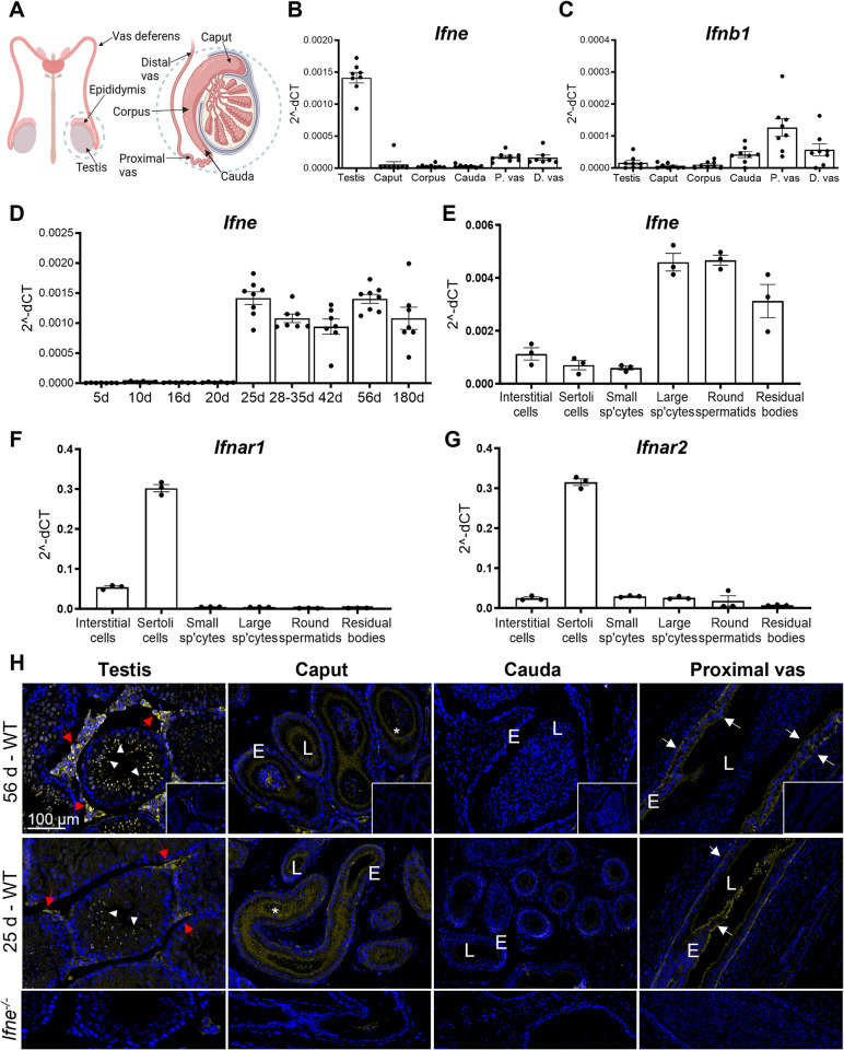
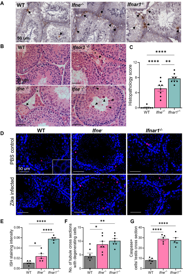
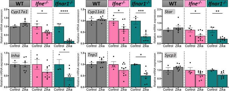
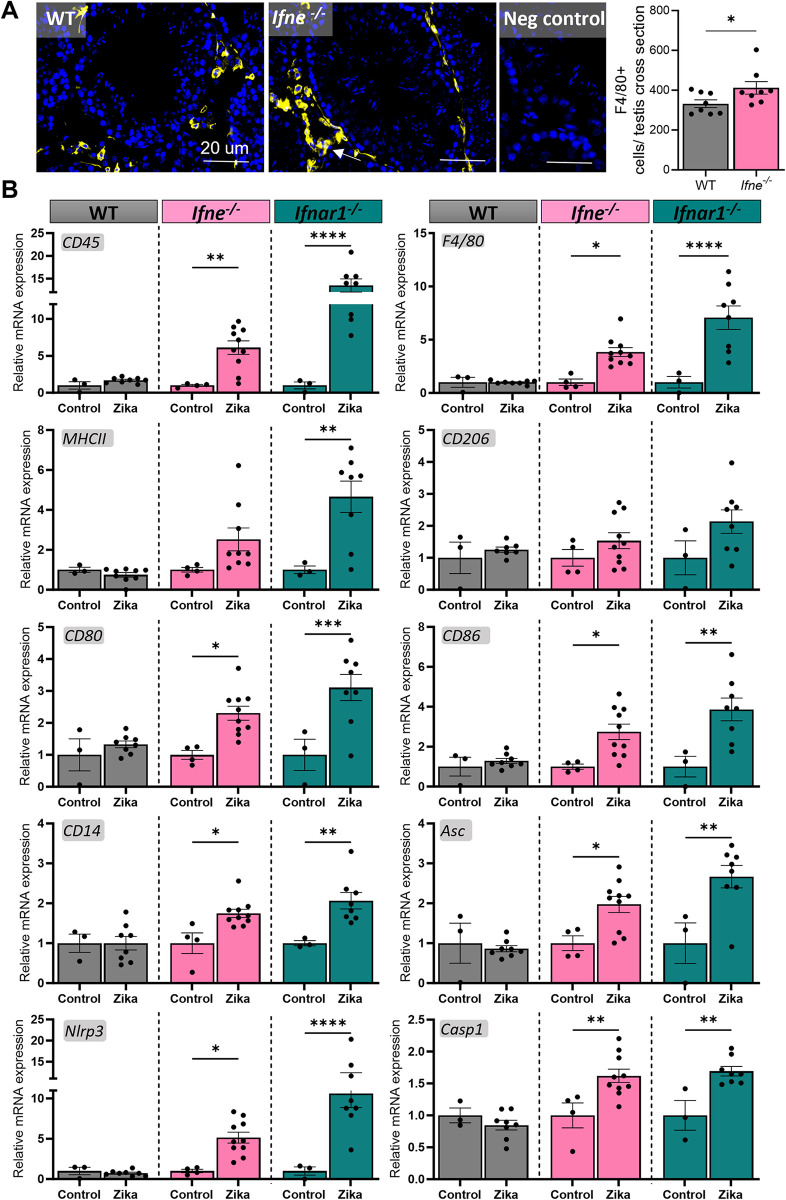
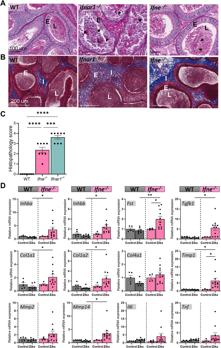
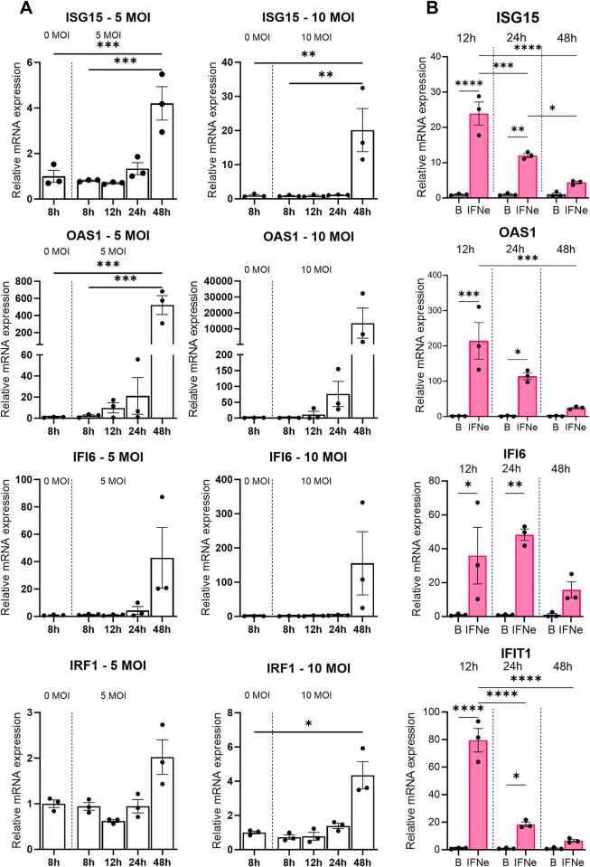
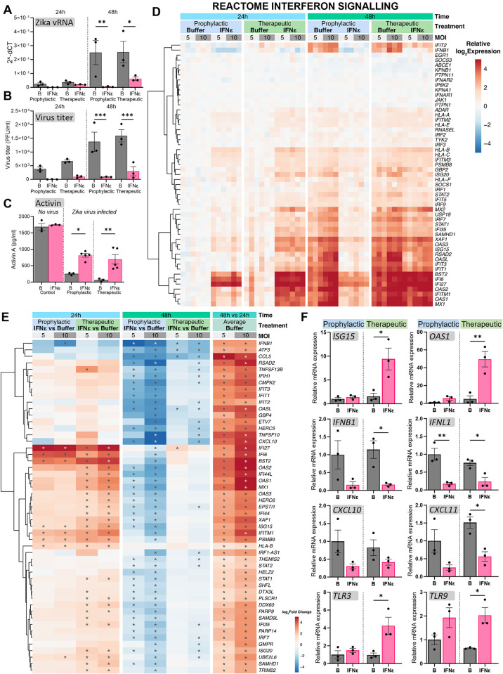
The testis is a reservoir for viruses that can cause persistent infection and adversely affect male reproductive health, an observation commonly attributed to deficiencies in inducible antiviral defence mechanisms. In this study, we demonstrate that interferon-epsilon (IFNε), a type I interferon initially discovered in female reproductive epithelia, is constitutively expressed by meiotic and post-meiotic spermatogenic cells, Leydig cells and macrophages in mouse testes. A similar distribution pattern was observed in human testes. Mice lacking IFNɛ were more susceptible to Zika virus-induced inflammation and damage of the testis and epididymis compared to wild-type mice. Exogenous IFNε treatment reduced the viral infection burden in cultured human testicular cells by inducing interferon-stimulated gene expression, and reducing inflammatory gene expression and cell damage. Treatment was more effective when administered prior to infection. These data indicate a critical role for constitutively-expressed IFNɛ in limiting viral infection and inflammatory damage in the male reproductive tract.


Conflict of interest statement

The authors have declared that no competing interests exist.

Figures

Fig 1
Fig 1. IFNɛ is constitutively expressed in the mouse male reproductive tract.
(A) Schematic diagram of the anatomy of the male reproductive tract. (B) Ifne and (C) Ifnb1 mRNA expression in the testis, epididymis (caput, corpus and cauda regions) and vas deferens (P. vas = proximal and D. vas = distal region of vas deferens) in 56-day-old mice (n = 8), (D) Ifne mRNA expression in the testis in mice aged 5–180 days (n = 6–8 per group). (E) Ifne and (F) type I interferon receptor Ifnar1 and (G) Ifnar2 mRNA expression in testicular cell types isolated from 44-day-old WT mice (n = 3 cell preps per cell type, Sp’cytes = spermatocytes). All mRNA expression data is relative to the reference gene Rplp0 (2^-dCt = relative copy number). (H) IFNε localisation in 56- and 25-day-old WT adult mouse male reproductive tract, compared to that of 56-day-old Ifne-/- mice using an anti-human IFNɛ antibody validated for mouse. Representative images from n = 4 mice. Inset: Isotype control. Yellow: IFNɛ. Blue: DAPI. Scale bar: 50 μm. Red arrows: Leydig cell and macrophage clusters. Arrowhead: post-meiotic spermatogenic cells. White arrows: Epithelial staining in vas deferens. Asterisk: stereocilia layer of the epididymal epithelium. E = epithelium of epididymis and vas deferens. L = lumen. The Biorender software was used to create Fig 1A.
Fig 2
Fig 2. IFNɛ is constitutively expressed in the human testis.
(A) IFNɛ localisation in the adult human testis, using an anti-human IFNɛ antibody at low and higher magnification; scale bars 50 and 20 μm respectively. Inset: Isotype control. Yellow: IFNɛ. Blue: DAPI. Arrows: Leydig cell and macrophage clusters. Arrowhead: post-meiotic spermatogenic cells embedded in Sertoli cell cytoplasm. Representative images from n = 3 testicular biopsy samples exhibiting normal testicular histology. (B) IFNE, IFNB1 and their receptors IFNAR1 and IFNAR2 mRNA expression measured by qRT-PCR in testicular tissue samples showing intact spermatogenesis without any signs of inflammation (Normal) compared with biopsies showing impaired spermatogenesis and focal immune cell infiltrates (I + WBC). n = 5–19 per group. Student’s t-test, ***P < 0.001. (C) Correlation analysis of IFNAR2 mRNA expression and the histological degree of immune cell infiltration in the testis biopsy site (scored from 0–4; 4 = dense, 3 = sparse, 2 = scattered, 1 = single cells, 0 = absent). n = 5–19 per group. Spearman’s coefficient method. (D) IFNɛ protein measured using a two-site ELISA assay in testicular interstitial fluid from three patient groups with different causes of infertility. MTA = Mixed testicular atrophy, SCO = Sertoli Cell Only phenotype, SA = spermatogenic arrest. LoD = Limit of detection: 3.9 pg/ml. Pooled samples, 25 μl testicular interstitial fluid per patient from 4 patients in each category.
Fig 3
Fig 3. IFNɛ-/- mice are more susceptible to epididymo-orchitis following Zika virus infection.
(A) Detection of Zika virus RNA (arrows) by in situ hybridization in the testis of adult Ifne-/- mice compared to C57BL/6 (WT) and Ifnar1-/- mice 7 days post-Zika infection. Scale bars = 50 μm, Representative images from testis sections of 5 mice per genotype. (B) Histopathological images of Zika infected Ifne-/- and Ifnar1-/- mouse testes compared to infected WT mice. Vascular congestion (arrows), oedema (asterisks) and degenerating germ cells (arrowheads). PAS stain, Scale bars: 20μm, representative images from n = 8. (C) Histopathological scoring of Zika infected WT, Ifne-/- and Ifnar1-/- mouse testes. (D) Apoptosis detection by immunofluorescence staining for Caspase 3/9 (pink) in Zika infected testes. Blue: DAPI. Scale bars = 50 μm. Inset: negative control with scale bar of 100 μm. n = 8. (E) Quantification of ISH staining intensity presented above in Fig 3A. (F) Quantification of the number of seminiferous tubule cross sections showing at least 1 degenerating spermatogenic cell per cross section in the testes of Zika infected mice. (G) Quantification of Caspase 3/9 positive cells per testis cross section presented in Fig 3D. (For C, E-G: n = 5–8, One-Way ANOVA, *p < 0.05, **p < 0.01, ****p < 0.0001).
Fig 4
Fig 4. Testicular functional genes are altered in IFNɛ-/- mice following 7 day Zika virus infection.
Gene transcripts related to steroidogenesis (Cyp11a1, Cyp17a1, Star), Sertoli cell function (Inha), and spermatogenesis (Tnp1, Sycp3) measured by qRT-PCR in testes from WT (grey bars), Ifne-/- (pink bars) and Ifnar1-/- (green bars) mice infected with Zika virus, compared to PBS injected controls. n = 3–10 per group. 2-Way ANOVA, *p < 0.05.
Fig 5
Fig 5. Ifne-/- mice show testicular immune infiltrates and NLRP3 inflammasome gene upregulation 7 days post-Zika virus infection.
(A) Immunofluorescence staining for F4/80+ immune cells (yellow) in the testes of Zika infected Ifne-/- and WT mice, and graph quantifying F4/80+ cells per testis cross section. Arrow: immune cell aggregates. Blue: DAPI. Scale bar: 20 μm. (B) Gene transcripts related to immune cells and the NLRP3 inflammasome measured by qRT-PCR in testes from WT (grey bars), Ifne-/- (pink bars) and Ifnar1-/- (green bars) mice infected with Zika virus, compared to PBS injected controls. (n = 8–10 per group. One-Way ANOVA for graph in Fig 5A, 2-Way ANOVA for 5B, *P < 0.05, **p < 0.01, ****p < 0.0001).
Fig 6
Fig 6. Ifne-/- mice develop epididymitis and subsequent fibrotic damage following 7-days post-Zika virus infection.
(A) Histopathological images of the epididymal corpus region of Zika infected Ifne-/- and Ifnar1-/- mice compared to infected WT mice. Degenerating germ cells prematurely released by the testis accumulating in the epididymal lumen (arrowheads), epithelial damage in Ifnar1-/- mice (arrows). E: epididymal epithelium. L: lumen. I: interstitial tissue. H&E stain. Scale bars: 100 μm. (B) Histopathology to assess fibrosis in the cauda region of the epididymis of Zika infected Ifne-/- and Ifnar1-/- and WT mice. Masson’s trichrome stain, Scale bars: 200 μm. (C) Histopathological scoring of damage to the epididymis of Zika infected Ifne-/- and Ifnar1-/- mice (n = 8 per group. One-Way ANOVA, ***p < 0.001, ****p < 0.0001). (D) Gene transcripts related to fibrosis and inflammation in the cauda region of Zika infected Ifne-/- mice compared to infected WT and uninfected Ifne-/- and WT mice, measured by qRT-PCR. n = 6–11 per group. Kruskal-Wallis test, *p < 0.05, **p<0.01, ***p< 0.001.
Fig 7
Fig 7. Zika virus infection induces most ISGs 48 hours post-infection in human Sertoli cells, while exogenous IFNɛ induces ISG production at an earlier time point.
(A) Interferon-stimulated gene (ISG) expression in human Sertoli cell cultures measured by qRT-PCR at 8, 12, 24 and 48 hours post-infection, with either 5 or 10 MOI Zika virus, compared to uninfected controls at 8 hours. (B) ISG signature induced by 100 IU exogenous IFNɛ treatment in uninfected human Sertoli cells, measured by qRT-PCR at 12, 24 and 48 hours post-treatment, compared to buffer treated-controls. All genes were normalised to the housekeeping gene RPLP0, B = Buffer treated cultures, IFNɛ = IFNɛ treated cultures. Each individual data point in the graphs represent the average of three technical replicates per culture round. One-Way ANOVA to compare more than 2 data sets, Student’s t-test to compare 2 data sets, *p < 0.05, **p<0.01, ***p< 0.001, ****p< 0.0001.
Fig 8
Fig 8. Treatment of human Sertoli cells with exogenous recombinant human IFNɛ has prophylactic and therapeutic benefits against Zika virus infection.
(A) qRT-PCR detection of Zika virus RNA (2^-dCt = relative copy number) and (B) infectious viral burden measured by plaque assays in primary human Sertoli cell cultures (HSerc) treated with 100 U/ml rhIFNɛ or buffer either before (prophylactic) or after (therapeutic) infection with 5 MOI Zika virus, assayed 24 and 48 hours post-infection. (C) Activin A protein levels produced by uninfected human Sertoli cells, compared Zika infected cultures treated with either buffer or prophylactic and therapeutic IFNɛ, measured by ELISA assay of culture media. (D) RNA-seq analysis of Sertoli cell cultures treated with rhIFNɛ or buffer prophylactically or therapeutically with 5 MOI (light grey) or 10 MOI (dark grey) Zika virus, assayed 24 and 48 hours post-infection. Heat map showing the relative log2 gene expression (compared to the prophylactically treated buffer controls at 24 hours post-infection at 5 MOI) for genes from the Reactome interferon signalling gene set. The scale is truncated at ±5. (E) Heat map showing genes significantly differentially expressed (with a twofold treat cut-off) for any IFNɛ treatment compared to its matched buffer control. Columns show log2 fold changes of IFNɛ versus buffer for each condition, as well as log2 fold changes for 48 hours versus 24 hours with buffer alone (i.e., the effect of virus over time; prophylactic and therapeutic treatments averaged). The scale is truncated at ±5. The asterisks indicate which genes were significantly changed for which comparisons. (F) qRT-PCR of anti-viral effectors, interferons, pro-inflammatory genes, and pattern recognition receptor genes from Sertoli cell cultures infected with 5 MOI Zika virus at 24 post-infection. B = Buffer treated cultures, IFNɛ = IFNɛ treated cultures. Each individual data point in the graphs represent the average of three technical replicates per culture round. One-Way ANOVA to assess data sets with one variable, 2-Way ANOVA for 2 variables. *P < 0.05, **P < 0.01, ***P < 0.001.

Similar articles

Cited by

References

    1. Le Tortorec A, Matusali G, Mahé D, Aubry F, Mazaud-Guittot S, Houzet L, Dejucq-Rainsford N. From Ancient to Emerging Infections: The Odyssey of Viruses in the Male Genital Tract. Physiological reviews (2020). 100, 1349–1414. doi: 10.1152/physrev.00021.2019 - DOI - PubMed
    1. Tsetsarkin KA, Acklin JA, Liu G, Kenney H, Teterina NL, Pletnev AG, Lim JK. Zika virus tropism during early infection of the testicular interstitium and its role in viral pathogenesis in the testes. PLOS Pathogens (2020). 16, e1008601. - PMC - PubMed
    1. Govero J, Esakky P, Scheaffer SM, Fernandez E, Drury A, Platt DJ, Gorman MJ, Richner JM, Caine EA, Salazar V., et al.. Zika virus infection damages the testes in mice. Nature (2016). 540, 438–442. doi: 10.1038/nature20556 - DOI - PMC - PubMed
    1. Duarte-Neto AN, Teixeira TA, Caldini EG, Kanamura CT, Gomes-Gouvêa MS, Dos Santos ABG, Monteiro RAA, Pinho JRR, Mauad T, da Silva LFF, et al.. Testicular pathology in fatal COVID-19: A descriptive autopsy study. Andrology (2022). 10, 13–23. doi: 10.1111/andr.13073 - DOI - PMC - PubMed
    1. Pilatz A, Arneth B, Kaiser R, Heger E, Pirkl M, Böttcher S, Fritzenwanker M, Renz H, Mankertz A, Schuppe HC, Wagenlehner F. Acute orchitis deciphered: Coxsackievirus B strains are the main etiology and their presence in semen is associated with acute inflammation and risk of persistent oligozoospermia. J Med Virol (2023). 95, e28970. doi: 10.1002/jmv.28970 - DOI - PubMed

Substances