. 2024 Dec 2;15(1):10486.
doi: 10.1038/s41467-024-54811-z.

Seipin governs phosphatidic acid homeostasis at the inner nuclear membrane

Affiliations

Seipin governs phosphatidic acid homeostasis at the inner nuclear membrane

Anete Romanauska et al. Nat Commun. .

Abstract

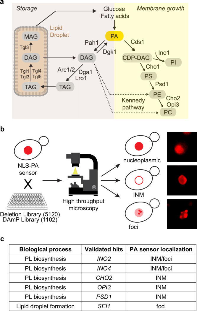
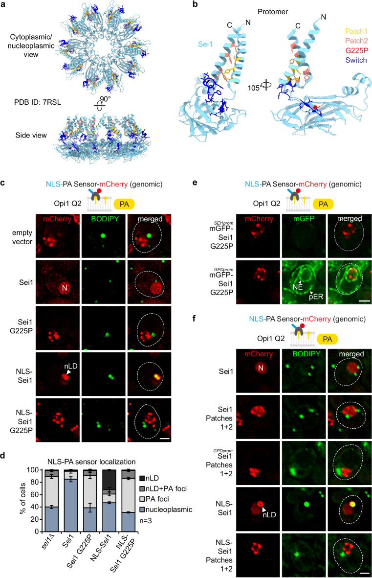
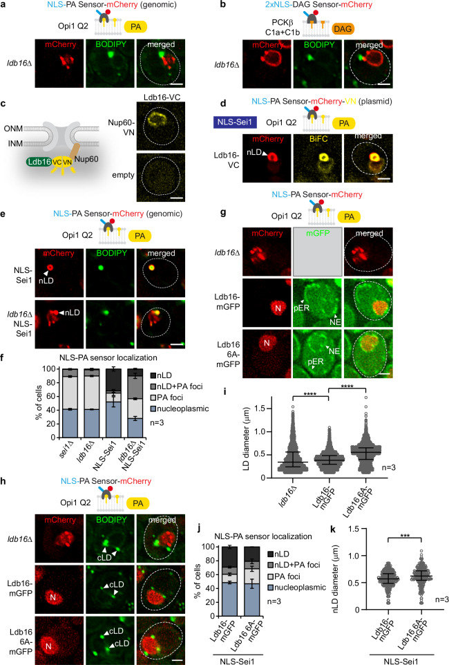
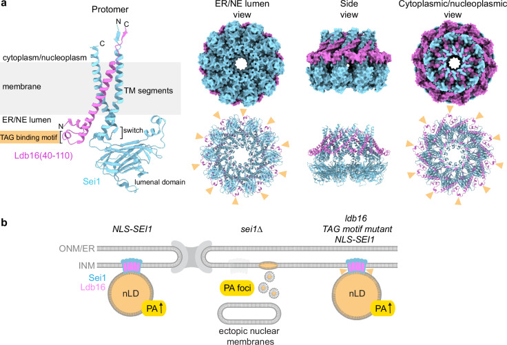
The nuclear envelope is a specialized subdomain of the endoplasmic reticulum and comprises the inner and outer nuclear membranes. Despite the crucial role of the inner nuclear membrane in genome regulation, its lipid metabolism remains poorly understood. Phosphatidic acid (PA) is essential for membrane growth as well as lipid storage. Using a genome-wide lipid biosensor screen in S. cerevisiae, we identify regulators of inner nuclear membrane PA homeostasis, including yeast Seipin, a known mediator of nuclear lipid droplet biogenesis. Here, we show that Seipin preserves nuclear envelope integrity by preventing its deformation and ectopic membrane formation. Mutations of specific regions of Seipin, some linked to human lipodystrophy, disrupt PA distribution at the inner nuclear membrane and nuclear lipid droplet formation. Investigating the Seipin co-factor Ldb16 reveals that a triacylglycerol binding site is crucial for lipid droplet formation, whereas PA regulation can be functionally separated. Our study highlights the potential of lipid biosensor screens for examining inner nuclear membrane lipid metabolism.


Conflict of interest statement

Competing interests: The authors declare no competing interests.

Figures

Fig. 1
Fig. 1. High-throughput screening uncovers PA regulators at the INM.
a Simplified scheme of yeast lipid biosynthesis depicting the two major branches leading to synthesis of phospholipids (Membrane growth) or triacylglycerol (TAG) (Storage). Phosphatidic acid (PA) is a central precursor. The Kennedy pathway (dashed line) channels diacylglycerol (DAG) into phospholipid production. CDP-DAG, cytidine diphosphate diacylglycerol; PS, phosphatidylserine; PE, phosphatidylethanolamine; PC, phosphatidylcholine; PI, phosphatidylinositol; MAG, monoacylglycerol. b Scheme of the high-throughput screening approach used to identify new regulators of PA at the INM with examples of phenotypes of PA localization observed using high-throughput screening. The 96-well plate icon was obtained from Clipground under a Creative Commons license. c Table showing validated hits, their biological function and PA sensor localization. Note that foci in ino2∆ and ino4∆ cells likely reflect nuclear lipid droplets. PL, phospholipid; INM, inner nuclear membrane.
Fig. 2
Fig. 2. Seipin regulates nuclear membrane architecture.
a Live imaging of wild-type or sei1∆ cells expressing a genomically integrated NLS-PA-mCherry sensor. BODIPY stains LDs. N, nucleus. Cell contours were marked with a dashed white line based on brightfield imaging. Scale bar, 2 μm. b Quantification of NLS-PA-mCherry sensor localization in (a). Mean value and standard deviation indicated. n, number of biological replicates. 480 cells for sei1∆ and 526 cells for wild-type analysed. Source data are provided as a Source Data file. c Analysis of NLS-PA-mCherry sensor foci co-localization with BODIPY in sei1∆ in (a). Mean value and standard deviation indicated. n, number of biological replicates. 591 foci in total analysed. Source data are provided as a Source Data file. d TEM analysis of a representative example of wild-type cell grown in SDC medium. N, nucleus; NE, nuclear envelope. Scale bar, 1 μm. e–h TEM analysis of representative examples of sei1∆ cells transformed with an empty vector and grown in SDC medium. Cells exhibit small lipid droplet-like structures marked with white asterisks in (e), intranuclear inclusions marked with a yellow arrowhead in (f), ectopic intranuclear membrane sheets marked with a red arrowhead in (g) and omega-shaped NE herniations marked with a green arrowhead in (h). See more TEM images in Supplementary Fig. 2. Insets show a magnified view of the marked areas. N, nucleus; NE, nuclear envelope; cLD, cytoplasmic lipid droplet; V, vacuole. Scale bar, 1 μm; 200 nm for insets. i Analysis of nuclear deformation frequency in nuclei of sei1∆ cells by TEM. sei1∆ cells were transformed with an empty vector and grown in SDC medium. Source data are provided as a Source Data file.
Fig. 3
Fig. 3. Seipin differentially alters lipid dynamics at the INM.
a Live imaging of wild-type or sei1∆ cells expressing plasmid-based 2xNLS-DAG-mCherry sensor and stained with BODIPY. INM, inner nuclear membrane. Scale bar, 2 μm. b Quantification of cells with 2xNLS-DAG-mCherry sensor foci in (a). Mean value and standard deviation indicated. n, number of biological replicates. 454 cells for sei1∆ and 628 cells for wild-type analysed. Source data are provided as a Source Data file. c Live imaging of wild-type, sei1∆ or cho1∆ cells expressing plasmid-based 3xNLS-PS-mCherry sensor and stained with BODIPY. INM, inner nuclear membrane; N, nucleus. cho1∆ cells were supplemented with ethanolamine and the sensor was expressed from the GPD promoter in cho1∆ cells. Scale bar, 2 μm. d Cartoon of the engineered NLS-Sei1 construct which contains the nuclear localization sequence (NLS) and the linker of the INM transmembrane protein Heh2 (aa93-317) appended to Sei1. Putative membrane topology of Sei1 is based on cryo-EM models. TEM analysis of a representative example of NLS-Sei1-expressing cells. Plasmid-based mGFP-NLS-SEI1 was expressed from the SEI1 promoter in a sei1∆ strain. N, nucleus; NE, nuclear envelope; nLD, nuclear lipid droplet. Asterisk marks a widened perinuclear space beneath an nLD. Scale bar, 0.5 μm. e Live imaging of sei1∆ cells expressing plasmid-based NLS-SEI1 and lipid sensors tagged with mCherry. Cells are stained with BODIPY. nLD, nuclear lipid droplet; INM, inner nuclear membrane. Scale bar, 2 μm. f Live imaging of sei1∆ cells expressing plasmid-based NLS-SEI1, NLS-PA-mGFP and lipid sensors tagged with mCherry. nLD, nuclear lipid droplet; INM, inner nuclear membrane. Scale bar, 2 μm.
Fig. 4
Fig. 4. Lipid droplet factors are localized to Sei1-formed nLDs.
a Live imaging of sei1∆ cells expressing plasmid-based NLS-SEI1, the indicated mGFP-tagged constructs and genomically integrated NLS-PA-mCherry sensor. All mGFP constructs were expressed from the GPD promoter, except PET10 (PET10 promoter) and PEX30 (TPI1 promoter). nLD, nuclear lipid droplet; cLD, cytoplasmic lipid droplet; pER, peripheral endoplasmic reticulum. Scale bar, 2  μm. b Live imaging of sei1∆ cells expressing plasmid-based NLS-SEI1, NLS-PA-mCherry-VN and the indicated VC-tagged constructs. All VC-tagged constructs were expressed from the GPD promoter, except PET10 (ADH1 promoter) and PEX30 (TPI1 promoter). nLD, nuclear lipid droplet. Scale bar, 2 μm. c Summary of Sei1-formed nLD proteome and lipidome.
Fig. 5
Fig. 5. PA defects at the INM in Sei1 lipodystrophy and TM contact mutants.
a Cartoon representation of S. cerevisiae Sei1 homodecamer (PDB ID: 7RSL) with select amino acid residues shown in atom (stick) representation. The amino acid residues, which were either substituted or deleted in corresponding Sei1 mutants, are colour-coded (yellow: Patch1, salmon: Patch2, red: G225P, blue: Switch). Note that the mutated amino acid residues do not map to the inter-subunit interfaces, and are therefore unlikely to disrupt the formation of the Sei1 ring assembly. b Close-up view of the Sei1 protomer (PDB ID: 7RSL, B conformation) shown in hybrid cartoon-atom representation as in Fig. 5a. c Live imaging of sei1∆ cells expressing genomically integrated NLS-PA-mCherry sensor and the indicated plasmid-based mGFP-SEI1 constructs. BODIPY stains LDs. nLDs have a BODIPY-positive core surrounded by a PA-rich shell. Note that even though cells contain mGFP-Sei1, the green BODIPY fluorescence signal is significantly brighter, hence Sei1 fluorescence remains undetectable when the settings for BODIPY imaging are applied. N, nucleus; nLD, nuclear lipid droplet. Scale bar, 2 μm. d Quantification of NLS-PA-mCherry sensor localization in (c). Mean value and standard deviation indicated. n, number of biological replicates. More than 435 cells analysed for each condition. Source data are provided as a Source Data file. e Live imaging of sei1∆ cells expressing genomically integrated NLS-PA-mCherry sensor and plasmid-based mGFP-sei1 G225P constructs from the endogenous SEI1 or a strong GPD promoter. Scale bar, 2 μm. f Live imaging of sei1∆ cells expressing genomically integrated NLS-PA-mCherry sensor and indicated plasmid-based mGFP-SEI1 constructs from the endogenous SEI1 or a strong GPD promoter. BODIPY stains LDs. N, nucleus; nLD, nuclear lipid droplet. Scale bar, 2 μm.
Fig. 6
Fig. 6. Ldb16’s hydroxyl residues govern proper cellular TAG accumulation but not PA metabolism at the INM.
a Live imaging of ldb16∆ cells expressing plasmid-based NLS-PA-mCherry sensor. BODIPY stains LDs. Scale bar, 2 μm. b Live imaging of ldb16∆ cells expressing plasmid-based 2xNLS-DAG-mCherry sensor and stained with BODIPY. Scale bar, 2 μm. c Experimental design for BiFC (bimolecular fluorescence complementation). VN, VC, complementary Venus fragments. Live imaging of wild-type cells expressing Ldb16 fused with VC and Nup60 fused with VN. Ldb16 is expressed from the GPD promoter. Nup60 is a basket nucleoporin, exclusively localized on the nuclear face of the nuclear pore complex. Empty vector is used as a control. Scale bar, 2 µm. d Live imaging of sei1∆ cells expressing plasmid-based NLS-PA-mCherry-VN, LDB16-VC and NLS-SEI1 constructs. Ldb16 is expressed from the GPD promoter. nLD, nuclear lipid droplet. Scale bar, 2 μm. e Live imaging of sei1∆ or sei1ldb16∆ cells expressing plasmid-based mGFP-NLS-SEI1 and genomically integrated NLS-PA-mCherry sensor. Cells are stained with BODIPY. nLD, nuclear lipid droplet. Scale bar, 2 μm. f Quantification of NLS-PA-mCherry sensor localization in (e). Mean value and standard deviation indicated. n, number of biological replicates. More than 380 cells analysed for each condition. Source data are provided as a Source Data file. g Live imaging of ldb16∆ cells expressing plasmid-based NLS-PA-mCherry sensor and indicated plasmid-based LDB16-mGFP constructs from the ADH1 promoter. N, nucleus. Scale bar, 2 μm. h Live imaging of ldb16∆ cells expressing plasmid-based NLS-PA-mCherry sensor and indicated plasmid-based LDB16-mGFP constructs expressed from the ADH1 promoter. BODIPY stains LDs. Note that even though cells contain Ldb16-mGFP, the green BODIPY fluorescence signal is significantly brighter, hence Ldb16 fluorescence remains undetectable when the settings for BODIPY imaging are applied. cLD, cytoplasmic lipid droplet. Scale bar, 2 μm. i Automated quantification of cellular LD diameter in (h). n, number of biological replicates. Median and interquartile range indicated. P value (****P  <  0.0001) determined by two-sided Kolmogorov-Smirnov test. Source data are provided as a Source Data file. j Quantification of NLS-PA-mCherry sensor localization in sei1ldb16∆ cells expressing genomically integrated NLS-PA-mCherry, plasmid-based mGFP-NLS-SEI1 and genomically integrated LDB16-mGFP constructs. LDB16 was expressed from the ADH1 promoter. Mean value and standard deviation indicated. n, number of biological replicates. 611 cells for LDB16-mGFP and 663 cells for ldb16 6A-mGFP were analysed. Source data are provided as a Source Data file. k Automated quantification of nLD diameter in sei1ldb16∆ cells expressing genomically integrated NLS-PA-mCherry, plasmid-based mGFP-NLS-SEI1 and genomically integrated LDB16-mGFP constructs. LDB16 was expressed from the ADH1 promoter. n, number of biological replicates. Median and interquartile range indicated. P value (***P  <  0.001) was determined by two-sided Kolmogorov-Smirnov test. 302 nLDs for LDB16-mGFP and 252 nLDs for ldb16 6A-mGFP were analysed. Source data are provided as a Source Data file.
Fig. 7
Fig. 7. Sei1 positions the TAG-binding domain of Ldb16 outside of the decameric ring.
a Cartoon representation of AlphaFold 3 model of S. cerevisiae Sei1·Ldb16(40-110) protomer (left), and both surface (right top) and cartoon (right bottom) representations of AlphaFold 3 model of Sei1·Ldb16(40-110) 10:10 ring assembly. The models are coloured by chain. Yellow arrowheads indicate putative serine/threonine-rich TAG binding motifs of Ldb16. For Sei1·Ldb16 prediction and confidence scores, see Supplementary Fig. 7. b A model illustrating the role of the Sei1-Ldb16 complex in regulating nLDs and INM lipid composition. When Sei1 is localized to the INM, nLDs with PA-enriched surfaces are formed. In the absence of Sei1, abnormal PA-rich but TAG-deficient droplets arise, accompanied by diverse defects in nuclear membrane structure. Mutations in the TAG-binding domain of Ldb16 (yellow arrowheads) reduce nLD numbers but slightly enlarge nLDs, suggesting impaired nLD biogenesis.

Similar articles

Cited by

References

    1. Bahmanyar, S. & Schlieker, C. Lipid and protein dynamics that shape nuclear envelope identity. Mol. Biol. Cell31, 1315–1323 (2020). - PMC - PubMed
    1. Mannino, P. J. & Lusk, C. P. Quality control mechanisms that protect nuclear envelope identity and function. J. Cell Biol.221, e202205123 (2022). - PMC - PubMed
    1. Ungricht, R. & Kutay, U. Mechanisms and functions of nuclear envelope remodelling. Nat. Rev. Mol. Cell Biol.10.1038/nrm.2016.153 (2017). - PubMed
    1. Harayama, T. & Riezman, H. Understanding the diversity of membrane lipid composition. Nat. Rev. Mol. Cell Biol.19, 281–296 (2018). - PubMed
    1. Carman, G. M. & Han, G. S. Regulation of phospholipid synthesis in the yeast Saccharomyces cerevisiae. Annu. Rev. Biochem.80, 859–883 (2011). - PMC - PubMed

Publication types

MeSH terms

Substances

LinkOut - more resources