. 2024 Dec 2;15(1):10465.
doi: 10.1038/s41467-024-54556-9.

EF-P and its paralog EfpL (YeiP) differentially control translation of proline-containing sequences

Affiliations

EF-P and its paralog EfpL (YeiP) differentially control translation of proline-containing sequences

Alina Sieber et al. Nat Commun. .

Abstract

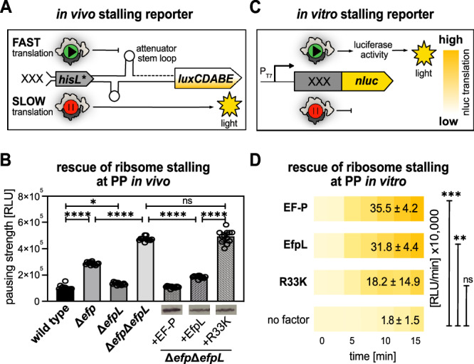
Polyproline sequences are deleterious to cells because they stall ribosomes. In bacteria, EF-P plays an important role in overcoming such polyproline sequence-induced ribosome stalling. Additionally, numerous bacteria possess an EF-P paralog called EfpL (also known as YeiP) of unknown function. Here, we functionally and structurally characterize EfpL from Escherichia coli and demonstrate its role in the translational stress response. Through ribosome profiling, we analyze the EfpL arrest motif spectrum and find additional sequences beyond the canonical polyproline motifs that both EF-P and EfpL can resolve. Notably, the two factors can also induce pauses. We further report that EfpL can sense the metabolic state of the cell via lysine acylation. Overall, our work characterizes the role of EfpL in ribosome rescue at proline-containing sequences, and provides evidence that co-occurrence of EF-P and EfpL is an evolutionary driver for higher bacterial growth rates.


Conflict of interest statement

Competing interests: The authors declare no competing interest.

Figures

Fig. 1
Fig. 1. Structural and phylogenetic analysis of the EfpL subgroup.
A Phylogenetic tree of EfpL (purple) and co-occurring EF-Ps (green). Colors of tip ends depict bacterial clades. Comparison of the KOW β3Ωβ4 loop in E. coli EF-P (taken from PDB: 6ENU; green) and EfpL (PDB: 8S8U, this study; purple). B Sequence logos of β3Ωβ4 loop of EfpL and co-occurring EF-Ps. C Comparison of structures of E. coli EF-P (taken from PDB: 6ENU) and EfpL (PDB: 8S8U, chain B, this study) with overall fold views and three domains.
Fig. 2
Fig. 2. The role of EfpL in bacterial physiology.
A Morphology analysis of E. coli BW23113 and isogenic mutant strains lacking efp (Δefp), efpL (ΔefpL), or both genes (ΔefpΔefpL). In strains overproducing EF-P (+EF-P), EfpL (+EfpL) and EfpL_R33K (+R33K) protein production was confirmed by immunoblotting utilizing the C-terminally attached His6-tag and Anti-His6 antibodies (α-His). Colony size was quantified by averaging the diameters (mean ∅ ± standard deviation (sd)) of 30 colonies on LB agar plates after 18 h of cultivation at 37 °C. Morphology analysis on plates was repeated two more times with similar results. B Doubling times (mean tD ± sd) were calculated from exponentially grown cells in LB (n ≥ 6, biological replicates). Statistically significant differences to wild-type growth according to two-way ANOVA test (P value (P) *P < 0.0332, ****P < 0.0001). C Growth analysis of E. coli cells in mixture over 72 h. An E. coli strain ΔcadC without any mutant growth phenotype under the test conditions was used as wild type. E. coli BW25113 ΔcadC was mixed with either E. coli BW25113 Δefp or ΔefpL and cultivated for 72 h. The share of the population was detected on LB agar plates (n = 4, biological replicates). Statistically significant differences to wild-type growth according to two-way ANOVA test (**P = 0.003, ****P < 0.0001). AC Source data are provided as a Source Data file.
Fig. 3
Fig. 3. The function of EfpL in alleviation of ribosome stalling.
A Scheme of the in vivo stalling reporter system. The system operates on the histidine biosynthesis operon of E. coli. In its natural form, the histidine biosynthesis gene cluster is controlled by the His-leader peptide (HisL), which comprises seven consecutive histidines. In our setup, the original histidine residues (His1 through His4) were replaced by artificial sequence motifs (XXX). Non-stalling sequences promote the formation of an attenuator stem loop (upper part) that impedes transcription of the downstream genes, thus ultimately preventing light emission. Conversely, in the presence of an arrest motif, ribosomes pause and hence an alternative stem loop is formed that does not attenuate transcription of the luxCDABE genes of Photorhabdus luminescens. B In vivo comparison of pausing at PPN in E. coli (for strain labeling and immunoblotting details see (A)). Pausing strength is given in relative light units (RLU) (n = 12, biological replicates, mean with sd indicated as error bars). Statistically significant differences according to an ordinary one-way ANOVA (*P < 0.0332, ****P < 0.0001, ns not significant). C Scheme of the in vitro cell-free stalling reporter assay. The system is based on nanoluc luciferase (nluc®) which is preceded by an artificial sequence motif (XXX). DNA is transcribed from a T7 promoter (PT7) using purified T7 polymerase (NEB). Pausing strength is proportional to light emission. D In vitro transcription and translation of the nLuc® variant nLuc_PPN. The absence (no factor) or presence of the respective translation elongation factors of E. coli (EF-P, EfpL, EfpL_R33K) is shown. Translational output was determined by measuring bioluminescence in a time course of 15 min and endpoints are given in relative light units (RLU/min±sd) (n ≥ 3, technical replicates). Statistically significant differences to the control (no factor) according to ordinary one-way ANOVA (**P = 0.0015, ***P = 0.0005, ns not significant). B, D Source data are provided as a Source Data file.
Fig. 4
Fig. 4. The target spectrum of EF-P and EfpL.
A Color code of the heat map corresponds to frequency of the motif to occur in pause site in the ribosome profiling analysis predicted with PausePred (From green to red = from low to high). First column: Top 29 motifs whose translation is dependent on EF-P and the control motif PAP in the ribosome profiling analysis comparing E. coli BW25113 with the efp deletion mutant (Δefp). Second column: Comparison of profiling data of Δefp and Δefp cells overexpressing efpLefp +EfpL) at these motifs. B In vivo comparison of rescue efficiency of a set of stalling motifs and the control PAP. Given is the quotient of relative light units measured in Δefp and corresponding trans-complementations by EF-P (+EF-P) and EfpL (+EfpL). Motifs are sorted according to pausing strength determined with our stalling reporter (n = 12, biological replicates, mean with sd indicated as error bars). C In vitro transcription and translation of nLuc® variants nLuc_3xRIPW (IPW) and nLuc_3xRPAP (PAP). The absence (no factor) or presence of the respective translation elongation factors of E. coli (EF-P/EfpL) is shown. Translational output was determined by measuring bioluminescence in a time course of 15 min and is given in relative light units measured at the end of the reaction (RLU/min ± sd) (n ≥ 3, technical replicates). Statistically significant differences to control (no factor) according to ordinary one-way ANOVA (****P < 0.0001, ns not significant). D Left part: Venn diagram of top 388 genes, whose translation depends on EF-P and EfpL. Dependency was determined by comparing asymmetry scores from genes encompassing top 29 stalling motifs listed in (A). Right part: Enriched protein classes to which EfpL-dependent genes belong. E Growth analysis of E. coli cells in mixture over 72 h in LB with 40 mM glucose. A ΔcadC strain without growth phenotype was used as the wild type. E. coli BW25113 ΔcadC was mixed with either E. coli BW25113 Δefp or ΔefpL. The share of the population was detected on LB agar plates (n = 4, biological replicates). Statistically significant differences to wild-type growth according to two-way ANOVA (***P = 0.0006, ns not significant). AC, E Source data are provided as a Source Data file.
Fig. 5
Fig. 5. EfpL acylation and its regulation in distinct bacteria.
A EfpL acylations according to refs. . Acylated lysines are depicted as part of a polypeptide, represented by the wavy line. B In vitro transcription and translation of the nLuc® variant nLuc_PPN. The absence (no factor) or presence of E. coli EF-P or EfpL and substitution variants EfpL_K23AcK, EfpL_K40AcK, EfpL_51AcK, EfpL_K57AcK is shown. Translational output was determined by measuring bioluminescence in a 15 min time course and is given in relative light units (RLU/min±sd) (n ≥ 3, technical replicates). Statistically significant differences according to ordinary one-way ANOVA (*P = 0.0364, ns not significant). C Growth analysis of E. coli BW25113 ΔefpΔuup trans-complemented with efp (+EF-P), efpL (+EfpL) or efpL substitution mutants (+EfpL_R33K/_K51R/_K51Q/_K51E) in M9-medium with 20 mM acetate as sole carbon source. Images of growth media were taken after 48 h (n = 3, biological replicates, mean with sd indicated as error bars). D Sequence logos of position 51 ± 3 amino acids in EfpL in Enterobacterales and Vibrionales. E In vivo comparison of pausing at PPN in E. coli Δefp cells and trans-complementations with EF-P/EfpL of E. coli (+EF-PEco/+EfpLEco), Yersinia enterocolitica (+EfpLYen), Serratia marcescens (+EfpLSma), P. luminescens (+EfpLPlu), Vibrio campbellii (+EfpLVca). Pausing strength is given in relative light units (RLU) (n = 6, biological replicates, mean with sd indicated as error bars). Statistically significant differences according to one-way ANOVA (*P = 0.0152, ***P = 0.0002, ns not significant). F In vitro analysis as in (B). The absence (no factor) or presence of elongation factors of E. coli (EF-PEco/EfpLEco) and V. campbellii (EfpLVca) is shown. (n ≥ 3, technical replicates) (statistics as in (B), ****P = 0.0001, **P = 0.0015, ***P = 0.0005). G Growth analysis of V. campbellii (in LM) and Vibrio natriegens (in LB) with corresponding deletions of efp (Δefp), efpL (ΔefpL), or both genes (ΔefpΔefpL) (n = 11; biological replicates, mean with sd indicated as error bars). H Phylogenetic analysis of predicted γ-proteobacterial growth rates comparing absence or presence of EfpL. Doubling times were predicted using codon usage bias in ribosomal proteins. (n = 786 genomes, median with top and bottom boundaries representing 1st and 3rd quartiles and whiskers indicating 1.5 times inter-quartile range). Statistically significant difference according to phylogenetic ANOVA (P = 0.029, P value based on 1000 permutations). B, C, EG Source data are provided as Source Data file.

References

    1. Tanner, D. R., Cariello, D. A., Woolstenhulme, C. J., Broadbent, M. A. & Buskirk, A. R. Genetic identification of nascent peptides that induce ribosome stalling. J. Biol. Chem.284, 34809–34818 (2009). - PMC - PubMed
    1. Doerfel, L. K. et al. EF-P is essential for rapid synthesis of proteins containing consecutive proline residues. Science339, 85–88 (2013). - PubMed
    1. Ude, S. et al. Translation elongation factor EF-P alleviates ribosome stalling at polyproline stretches. Science339, 82–85 (2013). - PubMed
    1. Hersch, S. J. et al. Divergent protein motifs direct elongation factor P-mediated translational regulation in Salmonella enterica and Escherichia coli. mBio4, e00180–00113 (2013). - PMC - PubMed
    1. Peil, L. et al. Distinct X/PP/X sequence motifs induce ribosome stalling, which is rescued by the translation elongation factor EF-P. Proc. Natl. Acad. Sci. USA110, 15265–15270 (2013). - PMC - PubMed

Publication types

MeSH terms

Associated data

LinkOut - more resources