. 2024 Dec 2;14(1):29949.
doi: 10.1038/s41598-024-80927-9.

TAp73 and ΔTAp73 isoforms show cell-type specific distributions and alterations in cancer

Affiliations

TAp73 and ΔTAp73 isoforms show cell-type specific distributions and alterations in cancer

Vaclav Hrabal et al. Sci Rep. .

Abstract

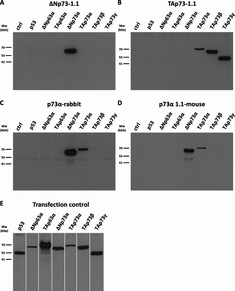
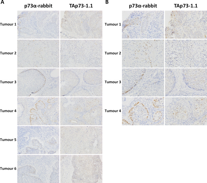
TP73 is a member of the TP53 gene family and produces N- and C-terminal protein isoforms through alternative promoters, alternative translation initiation and alternative splicing. Most notably, p73 protein isoforms may either contain a p53-like transactivation domain (TAp73 isoforms) or lack this domain (ΔTAp73 isoforms) and these variants have opposing or independent functions. To date, there is a lack of well-characterised isoform-specific p73 antibodies. Here, we produced polyclonal and monoclonal antibodies to N-terminal p73 variants and the C-terminal p73α isoform, the most common variant in human tissues. These reagents show that TAp73 is a marker of multiciliated epithelial cells, while ΔTAp73 is a marker of non-proliferative basal/reserve cells in squamous epithelium. We were unable to detect ΔNp73 variant proteins, in keeping with recent data that this is a minor form in human tissues. Most cervical squamous cell carcinomas (79%) express p73α, and the distribution of staining in basal cells correlated with lower tumour grade. TAp73 was found in 17% of these tumours, with a random distribution and no association with clinicopathological features. These data indicate roles for ΔTAp73 in maintaining a non-proliferative state of undifferentiated squamous epithelial cells and for TAp73 in the production of differentiated multiciliated cells.

Keywords: Cervical cancer; Endometrium; Fallopian tube; Multiciliated cells; Squamous epithelial stem cells; p73 isoforms.


Conflict of interest statement

Declarations. Competing interests: BV is a consultant for Moravian-Biotechnology spol. s r.o., who produced the antibodies used in this study. The company did not provide financial support for the study and had no influence on the design, execution or reporting of the work. All other authors report no conflicts of interest.

Figures

Fig. 1
Fig. 1
Schematic representation of TP63 and TP73 gene structure. (A) Transcription from promoter P1 and P2 of TP63 in combination with multiple possible 3ʹ-end splice variants may give rise to at least 10 mRNAs: TAp63α, TAp63β, TAp63γ, TAp63δ, TAp63ε, ΔNp63α, ΔNp63β, ΔNp63γ, ΔNp63δ, ΔNp63ε. (B) TP73 is more complex and 5ʹ-end variability is increased by additional splice variants Ex2, Ex2/3 and ΔNʹ. Alternative initiation of translation of transcripts at the first in-frame ATG of exon 4 may also result in another set of ΔTAp73 isoforms. There are also more 3ʹ-end splice variants: α,β,γ,δ,ε,ζ,η. TA1, TA1*, TA2, sequences important for transactivation. DBD DNA binding domain, OD oligomerisation domain, SAM sterile alpha motif, ID inhibitory domain.
Fig. 2
Fig. 2
Western blot analysis of antibody specificity. H1299 cells were transiently transfected with plasmids coding for p53, p63 isoforms or p73 isoforms, and non-transfected H1299 cells were used as a negative control (ctrl). (AD) Detection with antibodies recognising individual p73 isoforms: (A) ΔNp73-1.1 mouse monoclonal; (B) TAp73-1.1 recognising TAp73; (C) rabbit polyclonal p73α; (D) p73α-1.1 mouse monoclonal. Whole membranes are shown to reveal potential non-specific or cross-reactive bands. (E) To monitor successful transfection and protein expression, the membrane was cut into vertical strips representing each individual lane, and each lane was detected with antibodies that recognise the transfected protein. Original film images are shown in Supplementary Figure S1 online.
Fig. 3
Fig. 3
Immunocytochemical analysis of antibody specificity. Non-transfected H1299 cells or cells transfected with plasmids containing genes coding for p53 family isoforms (indicated down the left side of the figure) were formalin fixed and embedded in paraffin. Sections were immunocytochemically stained with the four antibodies indicated across the top of the figure (TAp73-1.1, affinity purified rabbit anti-p73α, mouse monoclonal p73α-1.1, and mouse monoclonal ΔNp73-1.1). Positive staining is seen as a brown precipitate and nuclei were counterstained with haemotoxylin (blue). Non-transfected H1299 cells were used as a negative control. Please note that transfections were performed transiently, therefore not all cells contain the expression plasmid.
Fig. 4
Fig. 4
Representation of antibody binding sites and binding motifs acquired from phage display epitope mapping. (A) Schematic representation and WebLogo image showing the TAp73-1.1 binding motif result from phage display epitope mapping. (B) Schematic representation of the binding sites of ΔNp73-1.1, ΔNp73-2.1 and ΔNp73-3.1. The WebLogo image shows the ΔNp73-1.1 binding motif from the phage display. (C) Binding site of p73α-1.1, p73α-2.1, p73α-3.1 and p73α rabbit antibodies.
Fig. 5
Fig. 5
Immunohistochemical staining of normal tissues. FFPE tissue sections were immunostained with p73α-1.1, TAp73-1.1 and ΔNp73-1.1 antibodies (brown) and nuclei were counterstained with haemotoxylin (blue). Additional tissues are shown in Supplementary Figure S3 online.
Fig. 6
Fig. 6
Triple immunofluorescent staining of p53, p63 and p73α in normal epithelial tissues. Formalin-fixed paraffin-embedded tissue sections were co-stained with antibodies to p73α (green), p63 (red) and p53 (yellow). Sections were counterstained with DAPI (blue). For easier visualisation, the DAPI staining channel was removed from the merged images on the right and the yellow channel of p53 was pseudo-coloured blue.
Fig. 7
Fig. 7
Immunohistochemical staining of p73α, ΔNp63 and Ki-67 in the indicated normal tissues. Positive staining is seen as brown reaction product. Nuclei are counterstained blue.
Fig. 8
Fig. 8
Immunohistochemical staining of cervical carcinoma. (A) Six different cervical tumours stained with affinity purified rabbit anti-p73α and mouse monoclonal TAp73-1.1 (brown). Sections were counterstained with haematoxylin (blue). (B) shows higher magnification images of the indicated tumours.

References

    1. Lane, D. P. & Crawford, L. V. T antigen is bound to a host protein in SV40-transformed cells. Nature278, 261–263 (1979). - PubMed
    1. Linzer, D. I., Maltzman, W. & Levine, A. J. The SV40 A gene product is required for the production of a 54,000 MW cellular tumor antigen. Virology98, 308–318 (1979). - PubMed
    1. Schmale, H. & Bamberger, C. A novel protein with strong homology to the tumor suppressor p53. Oncogene15, 1363–1367 (1997). - PubMed
    1. Kaghad, M. et al. Monoallelically expressed gene related to p53 at 1p36, a region frequently deleted in neuroblastoma and other human cancers. Cell90, 809–819 (1997). - PubMed
    1. Liu, Y., Su, Z., Tavana, O. & Gu, W. Understanding the complexity of p53 in a new era of tumor suppression. Cancer Cell.42, 946–967 (2024). - PMC - PubMed

MeSH terms

LinkOut - more resources