. 2024 Dec 2;8(1):276.
doi: 10.1038/s41698-024-00775-8.

Extreme wrinkling of the nuclear lamina is a morphological marker of cancer

Affiliations

Extreme wrinkling of the nuclear lamina is a morphological marker of cancer

Ting-Ching Wang et al. NPJ Precis Oncol. .

Abstract

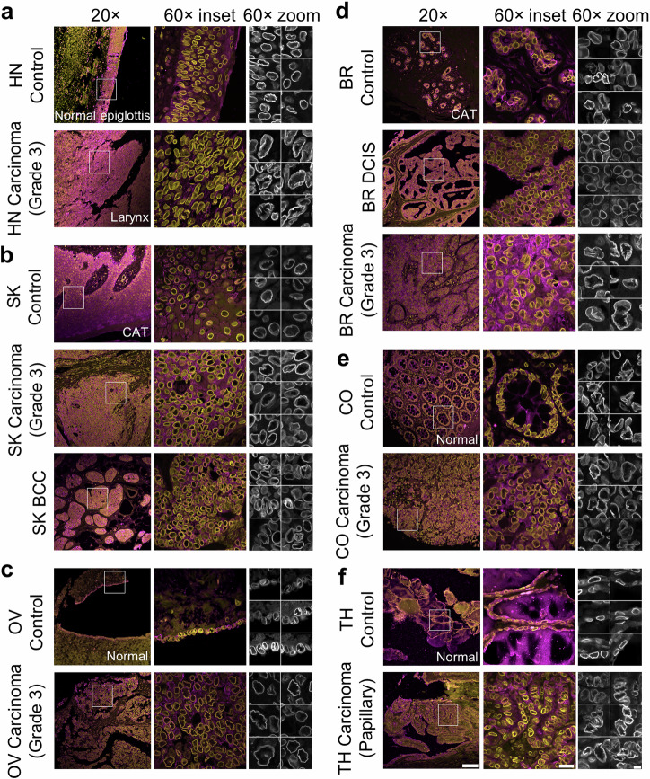
Nuclear atypia is a hallmark of cancer. A recent model posits that excess surface area, visible as folds/wrinkles in the lamina of a rounded nucleus, allows the nucleus to take on diverse shapes with little mechanical resistance. Whether this model is applicable to normal and cancer nuclei in human tissues is unclear. We image nuclear lamins in patient tissues and find: (a) nuclear laminar wrinkles are present in control and cancer tissue but are obscured in hematoxylin and eosin (H&E) images, (b) nuclei rarely have a smooth lamina, and (c) wrinkled nuclei assume diverse shapes. Deep learning reveals the presence of extreme nuclear laminar wrinkling in cancer tissues, which is confirmed by Fourier analysis. These data support a model in which excess surface area in the nuclear lamina enables nuclear shape diversity in vivo. Extreme laminar wrinkling is a marker of cancer, and imaging the lamina may benefit cancer diagnosis.


Conflict of interest statement

Competing interests: VCS is a consultant and equity holder for Femtovox Inc (unrelated to current work). TCW, CRD, SM, HP, SA, IS, VT, DGR, SC, and TPL declare no competing interests.

Figures

Fig. 1
Fig. 1. Nuclei in diverse control and cancer tissues have a wrinkled lamina.
a Formalin-fixed paraffin-embedded (FFPE) tissue stained for lamin B1 (yellow or gray) and pan-cytokeratin (magenta), imaged at 20× (left) and 60× (middle). The right columns show zoomed regions from the 60× image. Head and Neck (HN) tissue. b Skin (SK). CAT: Cancer Adjacent Tissue. c Ovarian (OV). d Breast (BR). e Colon (CO). f Thyroid (TH). Scale bars are 100, 20, and 5 µm for 20×, 60× inset, and 60× zoom, respectively.
Fig. 2
Fig. 2. Nuclear wrinkles are not an artifact of tissue processing and allow nuclei to assume diverse shapes.
a Excess perimeter distribution for pooled ductal carcinoma in situ (DCIS) patient nuclei and pooled breast cancer nuclei. Mean values were calculated from DCIS (n = 1882) and invasive cancer (n = 4575) nuclei. Error bars present standard deviations. b Collage of nuclei observed in formalin-fixed paraffin-embedded (FFPE) DCIS tissue sample (left) and invasive cancer samples (right) stained for lamin B1. c Frozen normal and cancer breast tissue fixed with acetone and stained for lamin B1. Scale bar is 5 µm. d Top: FFPE tissue section exhibiting shrinkage in the form of empty spaces (visible in brightfield) stained for lamin B1 (yellow) and pan-cytokeratin (magenta). Scale bar is 20 µm for 60× and 5 µm for zoomed. Below are zoomed regions from the lamin B1 image above (gray). e FFPE tissue sections featuring adjacent smooth and wrinkled nuclei. Scale bar is 5 µm. All images were taken at 60×.
Fig. 3
Fig. 3. Extreme nuclear wrinkling is a morphological feature of diverse cancers.
a Examples of nuclei sorted into classes 0–5 for deep learning model training. 0 = invalid nuclei, 1 = smooth, 2 = low frequency contour waviness, 3 = high frequency contour waviness, and 4 = inner wrinkles. b ResNet50 architecture of our model. An input image is processed through a 7×7 convolutional layer followed by max pooling. The network includes multiple residual learning blocks increasing in depth and complexity, each containing convolutional layers of varying filter sizes, with skip connections to ensure efficient training and feature extraction by mitigating the vanishing gradient problem. After feature extraction, we used an average pooling layer and a fully connected layer with a softmax function to classify the images into the five categories. c Bar plots showing the normalized corrected count of nuclei in each class. i. Head and neck; n = 1851, 2605, 1619, 1943, 1737 nuclei for adjacent, grades 1–3, respectively. ii. Skin; n = 848, 4180, 1637, 1487, 890 nuclei for adjacent, basal cell carcinoma (BCC), grades 1–3. iii. Ovary; n = 197, 3009, 2685, 10044, 1933, 3223, 4507 nuclei for control, mucinous, low grade serous, high grade serous, grades 1–3 endometrioid adenocarcinoma. iv. Breast; n = 3223, 2095, 859, 4296, 1054 nuclei for adjacent, ductal carcinoma in situ (DCIS), grades 1–3. v. Colon; n = 1458, 2147, 7011, 2521 nuclei for adjacent, grades 1–3. vi. Thyroid; n = 303, 10412 nuclei for normal and cancer, respectively. Error bars present a 95% confidence interval of the mean. *False discovery rate adjusted p < 0.05 by the Benjamini–Hochberg procedure.
Fig. 4
Fig. 4. Nuclear wrinkling and lymph node involvement.
Bar plots show the normalized corrected count of nuclei in each wrinkling class. i. Ovary; n = 134, 16944, 2414 nuclei for control, TNM grade N0, and N1, respectively. ii. Breast; n = 2541, 5124, 2489, 510 nuclei for control, N0–N2. iii. Colon; n = 1458, 7479, 2514, 1686 nuclei for control, N0–N2. iv. Thyroid; n = 303, 8395, 1601 nuclei for control, N0, and N1. Error bars present a 95% confidence interval of the mean. *False discovery rate adjusted p < 0.05 by the Benjamini–Hochberg procedure. Patient information is in Supplementary Table 3.
Fig. 5
Fig. 5. Elliptical Fourier analysis reveals higher contour irregularity in cancer.
Kernel density plots show the distribution of elliptical Fourier coefficient (EFC) ratios and nuclear areas for control and cancer tissues and for different tumor grades. The estimated kernel densities from the R = 100 random subsets were combined by averaging (see Methods). a Head and neck tissue; n = 1508, 1951, 1336, 250, 1603, 5140 nuclei for adjacent, grades 1–3, and pooled tumor grades, respectively. b Skin tissue; n = 762, 3724, 1658, 1509, 907, 4074 nuclei for adjacent, basal cell carcinoma (BCC), grades 1–3, and pooled grades. c Ovarian tissue; n = 58, 1530, 2027, 7085, 1382, 2074, 3251, 6707 nuclei for control, mucinous, low grade serous, high grade serous, grades 1–3 endometrioid adenocarcinoma, and pooled grades. d Breast tissue; n = 2455, 1918, 819, 3859, 1054, 5732 nuclei for adjacent, ductal carcinoma in situ (DCIS), grades 1–3, and pooled grades. e Colon tissue; n = 904, 1416, 5987, 2110, 9513 nuclei for adjacent, grades 1–3, and pooled grades. f Thyroid tissue; n = 69, 6030 nuclei for adjacent and cancer. p values for equality of means and homogeneity of scales obtained from the Kruskal-Wallis test and the Fligner test and adjusted using Benjamini-Hochberg false discovery rate corrections are labeled.

Similar articles

References

    1. Beale, L. Examination of sputum from a case of cancer of the pharynx and the adjacent parts. Arch. Med2, 1860–1861 (1860).
    1. Krishnamurti, U. G., Fitzgibbons, P. L., Connolly, J. L. Protocol for the Examination of Resection Specimens from Patients with Invasive Carcinoma of the Breast, https://www.cap.org/protocols-and-guidelines/cancer-reporting-tools/canc... (2023).
    1. Fitzgibbons, P. L., Connolly, J. L. Protocol for the Examination of Biopsy Specimens from Patients with Ductal Carcinoma In Situ (DCIS) of the Breasthttps://www.cap.org/protocols-and-guidelines/cancer-reporting-tools/canc... (2021). - PubMed
    1. Mete, O. Protocol for the Examination of Specimens From Patients With Carcinomas of the Thyroid Glan, https://www.cap.org/protocols-and-guidelines/cancer-reporting-tools/canc... (2023).
    1. Seethala, R. R. Protocol for the Examination of Specimens from Patients with Cutaneous Squamous Cell Carcinoma of the Head and Neck, https://www.cap.org/protocols-and-guidelines/cancer-reporting-tools/canc... (2022).

LinkOut - more resources