. 2025 Jan;44(2):413-436.
doi: 10.1038/s44318-024-00322-y. Epub 2024 Dec 2.

De novo variants in LRRC8C resulting in constitutive channel activation cause a human multisystem disorder

Affiliations

De novo variants in LRRC8C resulting in constitutive channel activation cause a human multisystem disorder

Mathieu Quinodoz et al. EMBO J. 2025 Jan.

Abstract

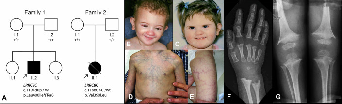
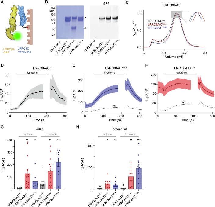
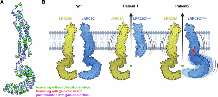
Volume-regulated anion channels (VRACs) are multimeric proteins composed of different paralogs of the LRRC8 family. They are activated in response to hypotonic swelling, but little is known about their specific functions. We studied two human individuals with the same congenital syndrome affecting blood vessels, brain, eyes, and bones. The LRRC8C gene harbored de novo variants in both patients, located in a region of the gene encoding the boundary between the pore and a cytoplasmic domain, which is depleted of sequence variations in control subjects. When studied by cryo-EM, both LRRC8C mutant proteins assembled as their wild-type counterparts, but showed increased flexibility, suggesting a destabilization of subunit interactions. When co-expressed with the obligatory LRRC8A subunit, the mutants exhibited enhanced activation, resulting in channel activity even at isotonic conditions in which wild-type channels are closed. We conclude that structural perturbations of LRRC8C impair channel gating and constitute the mechanistic basis of the dominant gain-of-function effect of these pathogenic variants. The pleiotropic phenotype of this novel clinical entity associated with monoallelic LRRC8C variants indicates the fundamental roles of VRACs in different tissues and organs.

Keywords: Channel Activation; Disease-causing Variants; Volume-regulated Anion Channels.


Conflict of interest statement

Disclosure and competing interests statement. The parents of the two patients in this study have been fully informed that the images shown would be used in a scientific research publication, and that the children will be fully identifiable in this publication, and given their consent. Showing such identifiable patient photographs is necessary for future clinical diagnosis in other affected children of the monoallelic variants of the LRRC8C gene similar to those reported here. We thankfully acknowledge the graciousness and generosity of the parents in giving this consent in the interest of helping future diagnoses in other affected children and in fostering knowledge about and research in this condition. The authors declare no competing interests.

Figures

Figure 1
Figure 1. Genetic and clinical features of the two cases with LRRC8C variants.
(A) Pedigree of patient P1 (Family 1, II.2), of Italian descent, and of patient P2 (Family 2, II.1), a Canadian individual whose parents are from Chile and Canada. The monoallelic de novo LRRC8C variants found in the patients are indicated below. (B, C) Facial appearance of P1 (at age 2 year) (B) and P2 (at age 2 years and 10 months) (C) showing short palpebral fissures, a small nose, and smooth facial features. Note the resemblance between the two unrelated individuals. (D, E) Prominent telangiectasia on the trunk (P1) and thigh (P2). (F, G) Radiographs of P1 showing thin diaphyses of the radius and ulna (F) and of the tibia and fibula (G) as well as marked metaphyseal fraying at the distal radius and ulna, distal femur and proximal tibia compatible with metaphyseal dysplasia. See statement in the disclosure and competing interest section at the end of the manuscript for parental consent to publishing these images. Source data are available online for this figure.
Figure 2
Figure 2. Characteristics of LRRC8C and of the variants identified.
(A) Schematic representation of the main LRRC8C transcript and its protein product. Both LRRC8Ctrunc (p.Leu400IlefssTer8) and LRRC8CV390L (p.Val390Leu), depicted by black arrowheads, fall into a region of the protein that is bereft of loss-of-function (pLoF, red track) or missense (orange track) variants in normal controls, as reported in the gnomAD database of normal controls, likely representing a sensitive site for DNA changes to result in pathogenic variants, as also shown by the MetaDome track (red, highest intolerance to changes; blue, lowest intolerance to changes). NTC N-terminal coil, TM transmembrane domain, EC extracellular loop, IC intracellular loop, LRRD leucine-rich repeat domain. (B) LRRC8C topology with secondary structure element of the pore PD indicated and the LRRD approximated as blue arc. (C) Conservation of the CTH2 and CTH3 region domains, with respect to the amino acid residues impacted by LRRC8Ctrunc and LRRC8CV390L, in human paralogues of LRRC8C (hLRRC8x) and orthologues from mouse (mLrrc8c) and zebrafish (zLrrc8c). LRRC8C is not present outside of bony vertebrates. (D) Model of the protein construct LRRC8Ctrunc based on the murine LRRC8CWT structure (PDBID: 8B40). The position of Leu 400 is indicated as a green sphere. (E) Structure of the murine LRRC8CWT subunit with the position of Val 390 indicated as red sphere. Inset (right) shows blowup of the region around Val 390 with its side chain represented as CPK model.
Figure 3
Figure 3. Structural properties of LRRC8C disease mutants.
(A) Size exclusion profiles of detergent-solubilized constructs of human LRRC8CWT and the two disease constructs LRRC8Ctrunc and LRRC8CV390L, each containing a C-terminal fusion to GFP. The elution of extracted proteins was detected via their GFP-fluorescence. (B, C) Structure of a homomeric hLRRC8Ctrunc construct. The cryo-EM densities at 7.3 Å prior to the application of symmetry (B) and at 4.9 Å after the application of C7 symmetry (C) are shown superimposed on a model of the PD of the heptameric protein viewed from within the membrane (top) and from the extracellular side (bottom, left). The density of the filter at 3.4 Å, obtained after masking, is shown on the bottom, right (C). Although the ESD is well-defined, the largest parts of the TM and the CSD are absent. (D) Cryo-EM density of a homomeric structure of hLRRC8CV390L at 4.4 Å processed without application of symmetry (C1). (E) Cryo-EM density of hLRRC8CWT at 3.7 Å processed without application of symmetry (C1). (D, E) Left, view from within the membrane contoured at 6.5 σ. Right, view from the extracellular side at 8.5 σ with model shown as ribbon and sidechains of the constricting residues Leu 105 as sticks. Bottom, right, view of the LRRDs from the cytoplasm at lower contour (3.5 σ). The relative positions of subunits within the heptamer are indicated. Source data are available online for this figure.
Figure 4
Figure 4. Heteromeric assembly and function of LRRC8C disease variants.
(A) Strategy for the investigation of heteromerization of LRRC8C disease variants. LRRC8C constructs carry an SBP tag for affinity purification and LRRC8A contains a fusion to GFP for detection. (B) Results from a single-step affinity purification. Left, Coomassie-stained SDS-PAGE gel showing both subunits. The LRRC8A-GFP and full-length LRRC8C constructs run at a similar molecular weight around 100 kDa (*), the LRRC8Ctrunc construct (<) at 50 kDa. Right, detection of in-gel fluorescence of GFP-tagged LRRC8A subunits demonstrates its pulldown by different LRRC8C constructs with similar efficiency. LRRC8AGFP refers to cells that were only transfected with LRRC8A-GFP as negative control. (C) SEC chromatogram of purified heteromeric LRRC8A/C channels containing WT and disease constructs of LRRC8C. Inset shows blowup of the peak region. (DF) Current densities (at 100 mV) of LRRC8−/− cells transfected with constructs coding for hLRRC8A and either hLRRC8CWT or disease mutants assayed by patch-clamp electrophysiology in the whole-cell configuration. Currents were periodically recorded by a ramp protocol. Perfusion with hypotonic medium is indicated by bar. Traces show mean and s.e.m. of (D), LRRC8A/CWT (n = 10), (E), LRRC8A/CV390L (n = 10), (F), LRRC8A/Ctrunc (n = 16) biological replicates recorded in buffer system 1 (Δsalt, 50 mM/kg gradient). (E, F) Mean current density of WT is shown as dashed line for comparison. (G, H) Mean current density (at 100 mV) in isotonic (measured 10 s after establishment of whole-cell configuration) and hypotonic conditions (measured 4 min after switch to hypotonic solutions) in (G), buffer system 1 (Δsalt) or (H), buffer system 2 (Δmannitol, 80 mM/kg gradient). Panels show measurements of individual cells (spheres), bars represent mean currents, errors are s.e.m. Asterisks indicate significant deviations from WT in the respective conditions in a Brown–Forsythe and Welch’s ANOVA test with Dunnett’s T3 multiple comparison testing *P < 0.05; **P < 0.01; ***P < 0.001 (Δsalt isotonic: LRRC8Ctrunc P = 0.0019, LRRC8CV390L P = 0.0406, Δsalt hypotonic: LRRC8Ctrunc P = 0.0041, LRRC8CV390L P = 0.0014, Δmannitol isotonic: LRRC8Ctrunc P = 0.012, LRRC8CV390L P = 0.008, Δmannitol hypotonic: LRRC8Ctrunc P = 0.0025, LRRC8CV390L P = 0.0009). The current increase upon exposure to hypotonic conditions in buffer system Δsalt was significant for LRRC8A/CWT (P = 0.035) and LRRC8A/CV390L (P = 0.0017) but not in case of LRRC8A/Ctrunc (P = 0.639). In buffer system Δmannitol, significant changes were observed for LRRC8A/CV390L (P = 0.0015) and LRRC8A/Ctrunc (P = 0.0296) but not LRRC8A/CWT (P = 0.721). Source data are available online for this figure.
Figure 5
Figure 5. Phenotype of human LRRC8C variants.
(A) Distribution of variants identified in human LRRC8C and their phenotypical consequence. Positions of variants are mapped on the LRRC8C subunit and shown as spheres. Variants leading to a premature termination of the protein without clinical phenotype (as ascertained in gnomAD, see text) are colored in green, the two variants discussed in this study with strong gain-of-function phenotype in red and violet. (B) Schematic depiction of the effect of disease variants in LRRC8C with pronounced gain-of-function phenotype on channel activity. Two subunits of a VRAC heteromer are shown, where the two LRRC8C disease mutants increase the flexibility of the subunit, leading to channel opening already under isotonic conditions.
Figure EV1
Figure EV1. Features of the hLRRC8C mutant V390L.
Comparison of the highest resolved cryo-EM maps (contoured at 8σ) obtained from the hLRRC8CV390L and hLRRC8CWT datasets. (A) Cryo-EM map of hLRRC8CV390L (top) and hLRRC8CWT (bottom) superimposed on indicated subunits. (B) hLRRC8CV390L (left) and hLRRC8CWT (right) densities viewed from the cytoplasm. (A, B) Parts of protein subunits that are defined in the density are shown as ribbons, subunit numbering is as in Fig. 3D, E.
Figure EV2
Figure EV2. Functional properties of hLRRC8C mutants in buffer set Δsalt.
(A) Representative current traces and current–voltage relationship of an LRRC8−/− cell transfected with constructs coding for hLRRC8A and hLRRC8CWT after swelling of cells in buffer set Δsalt showing the expected features of an LRRC8A/C channel. (B, C) Representative current traces and current–voltage relationships of an LRRC8−/− cell transfected with constructs coding for hLRRC8A and hLRRC8CV390L under isotonic (B) and hypertonic conditions (C). (D, E) Representative current traces and current–voltage relationships of an LRRC8−/− cell transfected with constructs coding for hLRRC8A and hLRRC8Ctrunc under isotonic (D) and hypertonic conditions (E). The similar general properties of currents obtained under isotonic and hypotonic conditions with respect to the negative reversal potential, the pronounced outward rectification and the inactivation at strongly positive voltages show characteristics of VRACs composed of LRRC8A and C-subunits. (AE) Currents were recorded by patch-clamp electrophysiology in the whole-cell configuration upon perfusion of cells with either isotonic (B, D) or hypotonic buffer (A, C, E, described in “Methods”). Normalized IV-plots are displayed (right, bottom). Voltage protocol is as shown in (A). (F) Reversal potentials of currents mediated by LRRC8A/Ctrunc or LRRC8A/CV390L in isotonic and hypotonic conditions of buffer system 1 (Δsalt) in comparison to LRRC8A/CWT currents measured in hypotonic conditions of the same buffer system. Cl refers to standard solutions, I and Glu to solutions where NaCl was replaced by equimolar amounts of NaI or NaGlu, respectively. Due to the lower anion concentration, reversal potentials are generally lower in hypotonic solutions. Panels show measurements of individual cells (spheres), bars represent mean currents, errors are s.e.m. Asterisks indicate significant deviations from WT in the respective conditions in a Brown–Forsythe and Welch’s ANOVA test with Dunnett’s T3 multiple comparison testing in case of the comparison of currents in hypotonic conditions and deviations from LRRC8Ctrunc in a unpaired t test with Welsch’s correction in case of isotonic conditions. Differences were usually not statistically significant (ns) except for one case (*P = 0.0375). (G) Inhibition of endogenous currents of LRRC8A/Ctrunc and LRRC8A/CV390L in response to perfusion of isotonic solutions of buffer condition Δsalt containing 50 µM DCPIB. Left, current decrease in representative traces in response to inhibitor addition measured at 100 mV by a ramp protocol. Dashed line refers to zero current. Right, fraction of inhibition of basal currents from respective constructs in response to DCPIB application. Measurements of individual cells are shown as spheres, bars represent mean currents, errors are s.e.m.
Figure EV3
Figure EV3. Functional properties of hLRRC8C mutants in buffer set Δmannitol.
(A, B) Mean current density (at 100 mV) of LRRC8A-/- cells (n = 10) (A) and HEK293 cells (n = 8) (B) recorded in buffer set Δmannitol. (C) Representative current traces and current–voltage relationship of LRRC8-/- cells transfected with constructs coding for hLRRC8A and hLRRC8CWT after swelling of cells in buffer set Δmannitol not leading to channel activation. (D) Mean current density (at 100 mV) of hLRRC8A/CWT channels (n = 12) recorded in buffer set Δmannitol. (E) Representative current traces and current–voltage relationship of LRRC8-/- cells transfected with constructs coding for hLRRC8A and hLRRC8CV390L after swelling of cells in buffer set Δmannitol. (F) Mean current density (at 100 mV) of hLRRC8A/CV390L (n = 11) recorded in buffer set Δmannitol. (G) Representative current traces and current–voltage relationship of LRRC8-/- cells transfected with constructs coding for hLRRC8A and hLRRC8Ctrunc after swelling of cells in buffer set Δmannitol. (H) Mean current density (at 100 mV) of hLRRC8A/Ctrunc channels (n = 10) recorded in buffer set Δmannitol. (AH) Currents were recorded by patch-clamp electrophysiology in the whole-cell configuration. Cannels were activated by perfusion of cells with hypotonic buffers (described in methods). (C, E, G) Normalized IV-plots are displayed (right, bottom). Voltage protocol is as shown in (C). (F, H) The mean current of WT is shown as dashed line for comparison. (D, F, H) Panels show mean of indicated number of biological replicates, errors are s.e.m. Note the different scale of the y-axis in the three panels.
Figure EV4
Figure EV4. Biochemical and functional characterization of the constructs LRRC8Ct379 and LRRC8Ct419.
(A) Models of truncated LRRC8 constructs. A ribbon representation of LRRC8Ctrunc is shown left as reference. The gray box indicates the region of interest in different constructs shown to the right: Center left, LRRC8Ct379, the region on the C-terminus of LRRC8Ctrunc (380–400) that is absent in this construct is highlighted in orange; Center right, LRRC8Ct419, the sequence extension compared to LRRC8Ctrunc (residues 401–419) is highlighted in green; Right, equivalent region of the LRRC8A mutant found in éburiffé mice (which is truncated after residue 442), with the extension compared to LRRC8Ctrunc highlighted in orange. (B) Fluorescence size exclusion chromatogram of GFP-fusions of the constructs LRRC8Ct379 and LRRC8Ct419 after extraction in detergent. Asterisk corresponds to the volume of the heptameric channel. While no oligomer is detected in case of the shorter LRRC8Ct379, the longer LRRC8Ct419 elutes at a similar volume as LRRC8Ctrunc. (C) Mean current density (at 100 mV) of LRRC8-/- cells transfected with constructs coding for LRRC8A and either LRRC8Ct419 (n = 5) or LRRC8Ct419 (n = 8) recorded by patch-clamp electrophysiology in the whole-cell configuration in isotonic (measured 10 s after establishment of whole-cell configuration) and hypotonic conditions (measured 4 min after switch to hypotonic solutions) in buffer system 1 (Δsalt). Panels show measurements of individual cells (spheres), bars represent mean currents, errors are s.e.m. Mean current densities obtained from LRRC8A/CWT (red) and LRRC8A/Ctrunc channels (blue, obtained from Fig. 4G) are shown as dashed line for comparison. Deviations of currents of both constructs to WT and mutated LRRC8C in the respective conditions were evaluated by a Brown–Forsythe and Welch’s ANOVA test with Dunnett’s T3 multiple comparison testing. Deviations between isotonic and hypotonic conditions for each construct was separately evaluated by unpaired t testing with Welsch’s correction applied. Differences to LRRC8A/CWT were in all cases found to be not statistically significant (LRRC8A/Ct379 isotonic, P = 0.542, hypotonic, P = 0.302; LRRC8A/Ct419 isotonic, P = 0.236, hypotonic, P = 0.366). Differences to LRRCA/Ctrunc were evaluated as significant (LRRC8A/Ct379 isotonic, P = 0.0023, hypotonic, P = 0.0028; LRRC8A/Ct419 isotonic, P = 0.0002, hypotonic, P = 0.0003). Current increases of both constructs upon change to hypotonic conditions were evaluated as statistically not significant (LRRC8A/Ct379 P = 0.274; LRRC8A/Ct419, P = 0.113).

References

    1. Afonine PV, Poon BK, Read RJ, Sobolev OV, Terwilliger TC, Urzhumtsev A, Adams PD (2018) Real-space refinement in PHENIX for cryo-EM and crystallography. Acta Crystallogr D Struct Biol 74:531–544 - PMC - PubMed
    1. Alghanem AF, Abello J, Maurer JM, Kumar A, Ta CM, Gunasekar SK, Fatima U, Kang C, Xie L, Adeola O et al (2021) The SWELL1-LRRC8 complex regulates endothelial AKT-eNOS signaling and vascular function. eLife 10:e61313 - PMC - PubMed
    1. Balkaya M, Dohare P, Chen S, Schober AL, Fidaleo AM, Nalwalk JW, Sah R, Mongin AA (2023) Conditional deletion of LRRC8A in the brain reduces stroke damage independently of swelling-activated glutamate release. iScience 26:106669 - PMC - PubMed
    1. Best L, Brown PD, Sener A, Malaisse WJ (2010) Electrical activity in pancreatic islet cells: The VRAC hypothesis. Islets 2:59–64 - PubMed
    1. Biesecker LG, Adam MP, Alkuraya FS, Amemiya AR, Bamshad MJ, Beck AE, Bennett JT, Bird LM, Carey JC, Chung B et al (2021) A dyadic approach to the delineation of diagnostic entities in clinical genomics. Am J Hum Genet 108:8–15 - PMC - PubMed

LinkOut - more resources