. 2024 Dec 3;14(1):30003.
doi: 10.1038/s41598-024-81773-5.

Low dose Adenoviral Vammin gene transfer induces myocardial angiogenesis and increases left ventricular ejection fraction in ischemic porcine heart

Affiliations

Low dose Adenoviral Vammin gene transfer induces myocardial angiogenesis and increases left ventricular ejection fraction in ischemic porcine heart

Niko Järveläinen et al. Sci Rep. .

Abstract

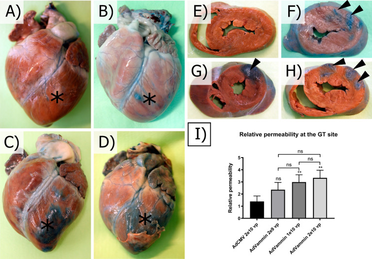
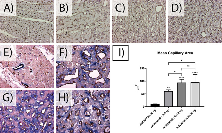
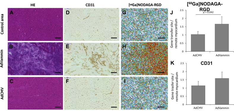
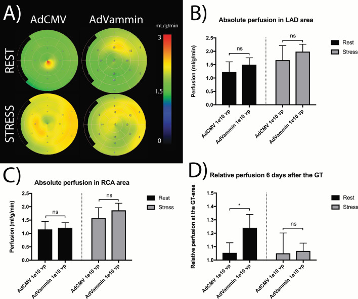
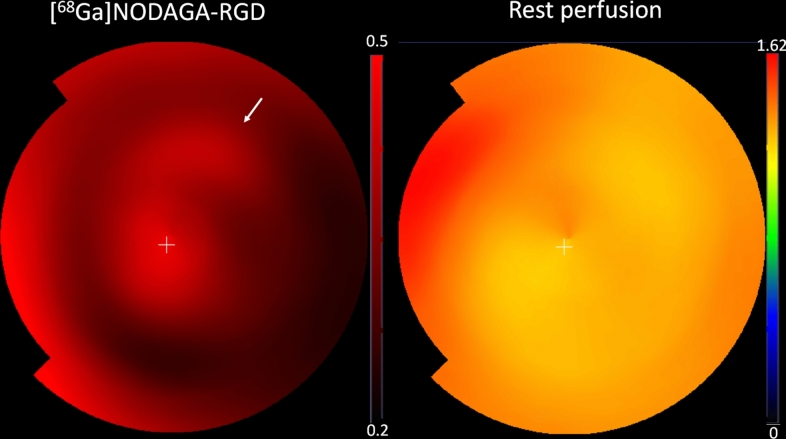
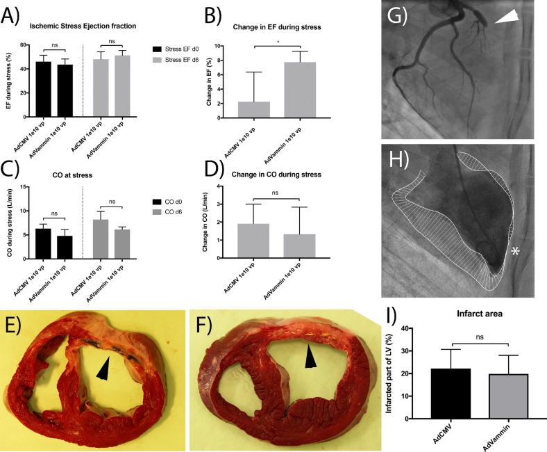
This preliminary study investigated if VEGFR-2 selective adenoviral Vammin (AdVammin) gene therapy could induce angiogenesis and increase perfusion in the healthy porcine myocardium. Also, we determined using a clinically relevant large animal model if AdVammin gene therapy could improve the function of a chronically ischemic heart. Low doses of AdVammin (dose range 2 × 10⁹-2 × 10¹⁰ vp) gene transfers were performed into the porcine myocardium using an endovascular injection catheter. AdCMV was used as a control. The porcine model of chronic myocardial ischemia was used in the ischemic studies. The AdVammin enlarged the mean capillary area and stimulated pericyte coverage in the target area 6 days after the gene transfers. Using positron emission tomography 15O-radiowater imaging, we demonstrated that AdVammin gene therapy increased perfusion in healthy myocardium at rest. AdVammin treatment also increased ejection fraction at stress in the ischemic heart, as detected using left ventricular cine angiography. In addition, we demonstrated successful in vivo imaging of enhanced angiogenesis using [68Ga]NODAGA-RGD peptide. However, AdVammin also increased tissue permeability and was associated with significant pericardial fluid accumulation, limiting AdVammin's therapeutic potential and emphasizing the importance of correct dosage.


Conflict of interest statement

Declarations. Competing interests: NJ is a shareholder of Saparo Translational Research Oy, a CRO for large animal studies. Ethical statement: The Finnish National Animal Experiment Board and the Animal Experiment Board of the University of Finland approved all animal experiments. All animal experiments were in compliance with the ARRIVE guidelines. People involved in animal work have Felasa B or C certification.

Figures

Fig. 1
Fig. 1
AdVammin gene transfer induced fluoroscopically visible injection sites in porcine myocardium six days after the gene transfer. (AD) shows representative angiograms of the left coronary artery six days after the gene transfer, taken from RAO 90 angle, for groups AdCMV 2 × 1010 vp (A), AdVammin 2 × 109 vp (B), 1 × 1010 vp (C), and 2 × 1010 vp (D). In the AdCMV group, no injection sites could be visualized (A). However, in the AdVammin treated animals, the injection sites (marked with arrowheads) are visible (BD). The effect was the most visible in the AdVammin 2 × 1010 vp dose (D). To highlight the effect, zoomed pictures of the angiograms for groups AdCMV 2 × 1010 vp and AdVammin 2 × 1010 vp are shown in J and E, respectively. In higher AdVammin doses, pericardial fluid accumulation six days after the gene transfer can be visualized in the fluoroscopy images (asterisk, C and D). The pericardial fluid accumulation for groups AdVammin 2 × 109 vp (G), 1 × 1010 vp (H), and 2 × 1010 vp (I) could also be visualized in echocardiography (asterisk, GI). No pericardial fluid was seen in the AdCMV control group (F). The left and right ventricle of the heart is marked in the echocardiography images by LV and RV, respectively. The echocardiograms (FI) represent the parasternal short-axis view of the heart. However, the views are suboptimal due to the limited acoustic window when imaging pigs with very narrow intercostal spaces.
Fig. 2
Fig. 2
AdVammin increased the vascular permeability six days after the gene transfer in the modified Miles Assay. Representative picture of the excised hearts (A-D) and cross-sections (EH) of the same heart for groups AdCMV 2 × 1010 vp (A and E), AdVammin 2 × 109 vp (B and F), AdVammin 1 × 1010 vp (C and G) and AdVammin 2 × 1010 vp (D and H) are shown in AH, with the target areas shown using black asterisks and arrowheads. The quantified relative vascular permeability in the gene transfer target site is represented in graph I. Statistical significance is marked by “ns” or asterisks (ns = p > 0.05 and ** = p ≤ 0.01). Increased vascular permeability was statistically significant in AdVammin 1 × 1010 vp and 2 × 1010 vp groups when compared to the AdCMV control group.
Fig. 3
Fig. 3
AdVammin gene transfer increased the mean capillary area in the porcine myocardium six days after the gene transfer. CD31-immunostained sections from the gene transfer area at 20 × magnification, with endothelial cells stained brown, for the AdCMV 2 × 1010 vp, AdVammin 2 × 109 vp, AdVammin 1 × 1010 vp and AdVammin 2 × 1010 vp groups are shown in A–D, respectively. The gene transfer with AdVammin significantly increased the mean capillary area in all dose groups. There was a significant difference in capillary size between the lowest AdVammin dosage and the higher dosages (I). Statistical significance is marked by “ns” or asterisks (ns = p > 0.05, * = p ≤ 0.05, ** = p ≤ 0.01, *** = p ≤ 0.001, **** = p ≤ 0.0001). Also, six days after the AdVammin gene transfer, the enlarged vessels showed clear pericyte coverage, as seen from the cd31-αSMA-immunostaining, with cd31 stained brown and αSMA stained blue (F–H). Representative pictures of CD31-αSMA-immunostained sections for groups AdCMV 2 × 1010 vp, AdVammin 2 × 109 vp, AdVammin 1 × 1010 vp, and AdVammin 2 × 1010 vp taken at 50 × magnification are shown in E–H, respectively.
Fig. 4
Fig. 4
Angiogenesis and myocardial [68 Ga]NODAGA-RGD-uptake in healthy porcine hearts. Compared with the remote myocardium (A and D) and the gene transfer site in the control group (AdCMV 1 × 1010 vp, C and F), there is a marked angiogenic response at the AdVammin gene transfer site (AdVammin 1 × 1010 vp, B and E). Blood vessel endothelium was detected by immunohistochemical staining with CD31 (D, E, F and K). Autoradiography shows increased [68 Ga]NODAGA-RGD uptake at the site of AdVammin gene transfer (H) as compared with the site of control gene transfer or the remote myocardium (G and I). The ratio of [68 Ga]NODAGA-RGD uptake in the gene transfer sites and the remote areas is significantly higher after AdVammin transfer than in the controls (J). Scale bars 100 µm (A-F) and 1000 µm (G-I). H&E = Hematoxylin and eosin.
Fig. 5
Fig. 5
AdVammin increased the relative perfusion at rest in the treatment areas six days after the gene transfer in healthy porcine hearts. The representative polar maps from the radio water-PET perfusion measurement are shown in A. The mean perfusion in the LAD and RCA areas corresponds to the treatment and control areas six days after the gene transfer for groups AdCMV 1 × 1010 vp and AdVammin 1 × 1010 vp are shown in B and C, respectively. The perfusion in the LAD area relative to the RCA area in rest was significantly increased in the AdVammin 1 × 1010 vp group compared to the AdCMV 1 × 1010 vp group (D). Statistical significance is marked by “ns” or asterisks (ns = p > 0.05 and * = p ≤ 0.05).
Fig. 6
Fig. 6
Representative image of [68 Ga]NODAGA-RGD peptide uptake and myocardial perfusion at rest. Figure shows polar maps of myocardial [68 Ga]NODAGA-RGD uptake 40–60 min after injection (standardized uptake value, SUV) and myocardial perfusion at rest (mL/min/g). Anterolateral wall (injection site) is marked with an arrow.
Fig. 7
Fig. 7
Effects of AdVammin gene transfer on left ventricle function and infarct size in a porcine chronic ischemia model. A stent obstructing blood flow was placed in the LAD (white arrowhead, G). The ejection fraction (EF) and cardiac output (CO) were assessed using a left ventricular cine angiography (H). There were no significant differences in EF or CO between AdCMV 1 × 1010 vp and AdVammin 1 × 1010 vp groups six days after the gene transfers (A and C). However, the change in EF six days after the treatment was significantly higher in the AdVammin 1 × 1010 vp group when compared to the AdCMV 1 × 1010 vp group (B). In CO, no differences were observed (D). The infarct areas were calculated from tetrazolium chloride incubated cross-sections of the excised hearts. Representative pictures for AdCMV 1 × 1010 vp and AdVammin 1 × 1010 groups are shown in E–F. There was no significant difference in the infarct size between the groups 28 days after the gene transfer (I). T. The dyskinesia in the anterior wall is seen in the left ventricular cine angiography (white asterisk, H).

References

    1. James, S. L. et al. Global, regional, and national incidence, prevalence, and years lived with disability for 354 Diseases and Injuries for 195 countries and territories, 1990–2017: A systematic analysis for the Global Burden of Disease Study 2017. The Lancet392, 1789–1858 (2018). - PMC - PubMed
    1. Wang, H. et al. Global, regional, and national life expectancy, all-cause mortality, and cause-specific mortality for 249 causes of death, 1980–2015: a systematic analysis for the Global Burden of Disease Study 2015. Lancet388, 1459 (2016). - PMC - PubMed
    1. Neumann, F. J. et al. 2018 ESC/EACTS Guidelines on myocardial revascularization. European Heart Journal vol. 40 87–165 Preprint at 10.1093/eurheartj/ehy394 (2019). - PubMed
    1. Neumann, F. J. et al. 2019 ESC Guidelines for the diagnosis and management of chronic coronary syndromes. European Heart Journal vol. 41 407–477 Preprint at 10.1093/eurheartj/ehz425 (2020). - PubMed
    1. Ylä-Herttuala, S., Bridges, C., Katz, M. G. & Korpisalo, P. Angiogenic gene therapy in cardiovascular diseases: Dream or vision? European Heart Journal vol. 38 1365–1371 Preprint at 10.1093/eurheartj/ehw547 (2017). - PMC - PubMed

LinkOut - more resources