. 2024 Dec 3;13(1):58.
doi: 10.1186/s40035-024-00452-7.

Enhanced prefrontal nicotinic signaling as evidence of active compensation in Alzheimer's disease models

Affiliations

Enhanced prefrontal nicotinic signaling as evidence of active compensation in Alzheimer's disease models

Saige K Power et al. Transl Neurodegener. .

Abstract

Background: Cognitive reserve allows for resilience to neuropathology, potentially through active compensation. Here, we examine ex vivo electrophysiological evidence for active compensation in Alzheimer's disease (AD) focusing on the cholinergic innervation of layer 6 in prefrontal cortex. Cholinergic pathways are vulnerable to neuropathology in AD and its preclinical models, and their modulation of deep layer prefrontal cortex is essential for attention and executive function.

Methods: We functionally interrogated cholinergic modulation of prefrontal layer 6 pyramidal neurons in two preclinical models: a compound transgenic AD mouse model that permits optogenetically-triggered release of endogenous acetylcholine and a transgenic AD rat model that closely recapitulates the human trajectory of AD. We then tested the impact of therapeutic interventions to further amplify the compensated responses and preserve the typical kinetic profile of cholinergic signaling.

Results: In two AD models, we found potentially compensatory upregulation of functional cholinergic responses above non-transgenic controls after onset of pathology. To identify the locus of this enhanced cholinergic signal, we dissected key pre- and post-synaptic components with pharmacological strategies. We identified a significant and selective increase in post-synaptic nicotinic receptor signalling on prefrontal cortical neurons. To probe the additional impact of therapeutic intervention on the adapted circuit, we tested cholinergic and nicotinic-selective pro-cognitive treatments. Inhibition of acetylcholinesterase further enhanced endogenous cholinergic responses but greatly distorted their kinetics. Positive allosteric modulation of nicotinic receptors, by contrast, enhanced endogenous cholinergic responses and retained their rapid kinetics.

Conclusions: We demonstrate that functional nicotinic upregulation occurs within the prefrontal cortex in two AD models. Promisingly, this nicotinic signal can be further enhanced while preserving its rapid kinetic signature. Taken together, our work suggests that compensatory mechanisms are active within the prefrontal cortex that can be harnessed by nicotinic receptor positive allosteric modulation, highlighting a new direction for cognitive treatment in AD neuropathology.

Keywords: Acetylcholine; Acetylcholinesterase inhibitor; Alzheimer’s disease; Cognitive reserve; Compensation; Electrophysiology; Nicotinic receptors; Optogenetics; Prefrontal cortex.


Conflict of interest statement

Declarations. Ethical approval: This manuscript contains animal research that was approved by the Faculty of Medicine Animal Care Committee at the University of Toronto in accordance with the guidelines of the Canadian Council on Animal Care (protocols #20011621, #20011658, #20011796). This work was conducted under the appropriate Material Transfer Authorizations. Competing interests: The authors have no conflicts or competing interests to declare.

Figures

Fig. 1
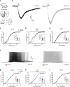
Fig. 1
Endogenous opto-ACh responses are upregulated in early to mid-AD. a.1 Image depicts compound transgenic mouse cross between TgCRND8 AD mouse model and ChAT-ChR2 mouse model expressing channelrhodopsin in cholinergic neurons. Image depicts coronal brain section from these mice, showing the recording electrode positioned over layer 6 of the prefrontal cortex. a.2 Schematic adapted from Power et al., 2023, represents the cholinergic prefrontal synapse receiving optogenetic stimulus (0.5-s pulse train of decreasing frequency). Afferents release endogenous acetylcholine (ACh) which binds to nicotinic and muscarinic receptors, exciting postsynaptic pyramidal neurons. b Representative opto-ACh responses from non-transgenic (nTg) control (black) and TgCRND8 AD (grey) neurons measured in voltage clamp (Vm =  − 75 mV). c, d Graphs show significant increases in rise speed (P < 0.01) (c) and peak amplitude (P < 0.01) (d) of opto-ACh currents in TgCRND8 early AD responses relative to age-matched controls (3–6 months old). e Representative opto-ACh responses from WT control (black) and TgCRND8 AD (grey) neurons measured in current clamp. f, g Graphs show significant increase in firing rise (P < 0.0001) (f) and peak firing frequency (P < 0.05) (g) of opto-ACh firing responses in TgCRND8 early/mid AD responses relative to age-matched controls. Mice aged 3 to 6 months (nTg: 4.5 ± 0.4 months, n = 10 animals; 3–4 brain slices per animal, TgCRND8: 4.8 ± 0.3 months, n = 10 animals, 3–4 brain slices per animal)
Fig. 2
Fig. 2
Cholinergic signaling did not differ from non-transgenic in late AD. a Representative opto-ACh responses from non-transgenic (nTg) control (black) and TgCRND8 AD (grey) neurons at late stage of disease measured in voltage clamp (Vm =  − 75 mV). b, c Graphs show no difference in rising speed (b) and peak amplitude (c) of opto-ACh currents in TgCRND8 late-AD responses compared to age-matched controls. d Representative opto-ACh responses from WT control (black) and TgCRND8 AD (grey) neurons at late stage of disease measured in current clamp. e, f Graphs show no difference in firing rise (e) and peak firing frequency (f) of opto-ACh firing responses in TgCRND8 late-AD responses compared to age-matched controls. Mice aged 7–12 months (nTg: 9.3 ± 0.5 months, n = 10 animals, 3–4 brain slices per animal, TgCRND8: 10.1 ± 0.6 months, n = 10 animals, 3–4 brain slices per animal)
Fig. 3
Fig. 3
Early cholinergic upregulation conserved in different species and AD models. a Image depicts TgF344 AD rat model and a coronal brain section with the recording electrode positioned over layer 6 of the prefrontal cortex. b Representative responses to exogenous bath-applied acetylcholine from F344 non-transgenic (nTg) control (black) and TgF344 AD (grey) neurons measured in voltage clamp (Vm =  − 75 mV). ce Graphs show that the cumulative frequency of peak amplitude of acetylcholine current was not different at 8 months (c), was significantly higher for TgF344 AD responses than controls at 12 months (P < 0.01) (d), and was not different at 18 months (e). Insets show the data in scattergram. f Representative responses to exogenous bath-applied acetylcholine from F344 WT control (black) and TgF344 AD (grey) neurons measured in current clamp. g, h Graphs show that the cumulative frequency of time to peak firing of acetylcholine response was not different at 8 months (g), was significantly faster for TgF344 AD responses than controls at 12 months (P < 0.01) (h), and was not different at 18 months (i). Insets show the data in scattergram. Rats aged in three cohorts: 8 months (nTg: 8.5 ± 0.2 months, n = 12 animals, 4–5 brain slices per animal, TgF344: 8.4 ± 0.2, n = 9 animals, 4–5 brain slices per animal), 12 months (nTg: 12.4 ± 0.2 months, n = 13, 4–5 brain slices per animal, TgF344 12.5 ± 0.2 months, n = 10 animals, 4–5 brain slices per animal), and 18 months (nTg: 17.7 ± 0.3 months, n = 14 animals, 4 –5 brain slices per animal, TgF344: 17.6 ± 0.2 months, n = 12 animals, 4–5 brain slices per animal)
Fig. 4
Fig. 4
Early cholinergic upregulation in the AD model is specific to nicotinic receptor signalling. a Schematic depicts a cholinergic synapse in the presence of nicotinic receptor antagonist DHβE. b Representative opto-ACh responses from non-transgenic (nTg) control (black) and TgCRND8 AD (grey) neurons measured in voltage clamp (Vm =  − 75 mV) before and after DHβE. c, d Graphs show that DHβE caused greater declines in rise speed (P < 0.05) (c) and peak amplitude (P < 0.05) (d) of opto-ACh currents in TgCRND8 AD responses relative to controls (age range: 4.5 to 12.2 months, mean = 7.9 ± 0.7 months, n = 14 animals, 1–3 brain slices per animal). e Schematic depicts a cholinergic synapse in the presence of broad muscarinic receptor antagonist atropine. f Representative opto-ACh responses from non-transgenic (nTg) control (black) and TgCRND8 AD (grey) neurons measured in voltage clamp before and after atropine. Graphs show that atropine elicited changes in rising speed (g) and peak amplitude (h) that were not different between TgCRND8 AD and control responses (age range: 3.1 to 12.2 months, mean = 6.4 ± 0.7 months, n = 20 animals, 1–3 brain slices per animal). i Schematic depicts a cholinergic synapse in the presence of muscarinic M2 receptor antagonist AF-DX 116. j Representative opto-ACh responses from non-transgenic (nTg) control (black) and TgCRND8 AD (grey) neurons measured in voltage clamp before and after AF-DX 116. Graphs show that AF-DX 116 elicited changes in rising speed (k) and peak amplitude (i) that were not different between TgCRND8 AD and control responses (age range: 4.2 to 12.2 months, mean = 7.8 ± 0.9 months, n = 13 animals, 1–3 brain slices per animal)
Fig. 5
Fig. 5
Galantamine and a novel nicotinic treatment strategy further enhance cholinergic upregulation in AD model. a Schematic depicts a cholinergic synapse in the presence of acetylcholinesterase inhibitor galantamine. Galantamine slowed the degradation of acetylcholine, prolonging its availability at the synapse. b Representative opto-ACh responses from a TgCRND8 AD neuron before (grey) and after galantamine (purple) treatment. ce Graphs show that galantamine elicited no change in the rising speed (c) but significant increases in peak amplitude (P < 0.01) (d) and response duration (e), as measured by the decay constant (P < 0.01) in TgCRND8 AD responses (age range 3–6 months: mean = 5.2 ± 0.6 months, n = 5 animals, 1–3 brain slices per animal). f Schematic depicts the activation of postsynaptic nicotinic receptors by acetylcholine and the action of nicotinic positive allosteric modulator NS9283 in increasing nicotinic receptor conductance. g Representative opto-ACh responses from a TgCRND8 AD neuron before (grey) and after (blue) NS9283 treatment. hj Graphs show that NS9283 elicited increases in rise speed (P < 0.05) (h) and peak amplitude (P < 0.001) (i) with no change in the response duration (j) (age range 3–6 months: mean = 4.7 ± 0.4 months, n = 6 animals, 1–3 brain slices per animal)

References

    1. Stern Y. Cognitive reserve in ageing and Alzheimer’s disease. Lancet Neurol. 2012;11:1006–12. - PMC - PubMed
    1. Valenzuela MJ, Sachdev P. Brain reserve and dementia: a systematic review. Psychol Med. 2006;36:441–54. - PubMed
    1. Stern Y, Gurland B, Tatemichi TK, Tang MX, Wilder D, Mayeux R. Influence of education and occupation on the incidence of Alzheimer’s disease. JAMA. 1994;271:1004–10. - PubMed
    1. Scarmeas N, Levy G, Tang M-X, Manly J, Stern Y. Influence of leisure activity on the incidence of Alzheimer’s disease. Neurology. 2001;57:2236–42. - PMC - PubMed
    1. Stern Y, Alexander GE, Prohovnik I, Mayeux R. Inverse relationship between education and parietotemporal perfusion deficit in Alzheimer’s disease. Ann Neurol. 1992;32:371–5. - PubMed

Publication types

LinkOut - more resources