. 2024 Dec 2;22(1):747.
doi: 10.1186/s12951-024-03019-w.

Engineering extracellular vesicles to transiently permeabilize the blood-brain barrier

Affiliations

Engineering extracellular vesicles to transiently permeabilize the blood-brain barrier

Francesca Tomatis et al. J Nanobiotechnology. .

Abstract

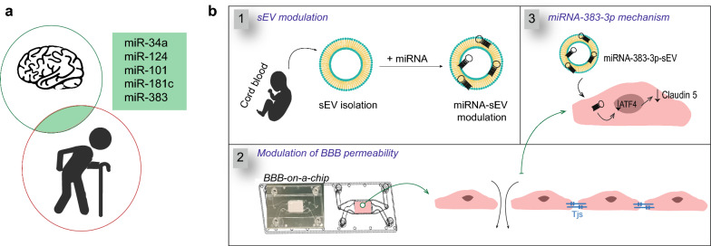
Background: Drug delivery to the brain is challenging due to the restrict permeability of the blood brain barrier (BBB). Recent studies indicate that BBB permeability increases over time during physiological aging likely due to factors (including extracellular vesicles (EVs)) that exist in the bloodstream. Therefore, inspiration can be taken from aging to develop new strategies for the transient opening of the BBB for drug delivery to the brain.

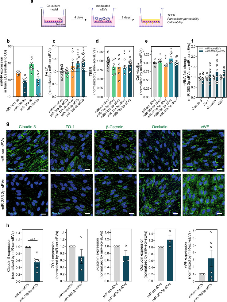
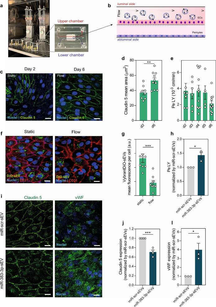
Results: Here, we evaluated the impact of small EVs (sEVs) enriched with microRNAs (miRNAs) overexpressed during aging, with the capacity to interfere transiently with the BBB. Initially, we investigated whether the miRNAs were overexpressed in sEVs collected from plasma of aged individuals. Next, we evaluated the opening properties of the miRNA-enriched sEVs in a static or dynamic (under flow) human in vitro BBB model. Our results showed that miR-383-3p-enriched sEVs significantly increased BBB permeability in a reversible manner by decreasing the expression of claudin 5, an important tight junction protein of brain endothelial cells (BECs) of the BBB, mediated in part by the knockdown of activating transcription factor 4 (ATF4).

Conclusions: Our findings suggest that engineered sEVs have potential as a strategy for the temporary BBB opening, making it easier for drugs to reach the brain when injected into the bloodstream.

Keywords: Blood–brain barrier; Claudin 5; Extracellular vesicles; MicroRNA; Microfluidic system; Modulation.


Conflict of interest statement

Declarations. Ethics approval and consent to participate: The collection of umbilical cord blood (UCB) was approved by the ethical committee of Dr. Daniel de Matos Maternity Hospital in Coimbra (protocol HUC-01-11), Portugal. The collection of adult blood was approved by the Faculty of Medicine of the University of Coimbra and the Portuguese Institute for Blood and Transplantation (IPST) in the facilities of Blood and Transplantation Center of Coimbra (CSTC) (protocol: CE-088/2020). All the donors of the UCB and adult blood signed an informed consent form, in compliance with the Portuguese legislation. Consent for publication: Not applicable. Competing interests: The authors declare no competing interests.

Figures

Fig. 1
Fig. 1
Engineering extracellular vesicles to permeabilize the BBB. a Identification of the miRNA candidates that are simultaneously altered during aging or age-related diseases and have shown to affect BBB permeability. b Overview of the experimental setup. b.1 sEVs collected from human UCB plasma were transfected with a miRNA of interest by a chemical agent. b.2 MiRNA-enriched sEVs were tested in human in vitro BBB models both in static and flow conditions. b.3 Identification of the mechanism behind the transient opening of the BBB after exposure to miRNA-enriched sEVs
Fig. 2
Fig. 2
Expression of miRNAs in sEVs changes during aging. a Scheme representing the list of miRNAs (in bold) selected for our study. b Table with the ages of the adult plasma donors (expressed in years old). For both males and females, the average is 61.6 years old. c RT-qPCR analyses performed to characterize the miRNA content in sEVs from plasma of aged donors (aged 58–64; labeled as “old sEVs”) compared to the sEVs from umbilical cord blood (UCB, labeled as “young sEVs”). Five miRNAs were evaluated. The data are expressed as mean ± SEM, n = 5 biological samples (2–3 technical replicates per biological sample). The unpaired t-test statistical analysis was performed for each miRNA, comparing the young sEVs with old sEVs from male and female donors. The * means p < 0.05, ** means p < 0.01, *** means p < 0.001
Fig. 3
Fig. 3
Modulation of sEVs with miRNAs. a Schematic representation of the sEV modulation protocol. The loading of miRNAs on the sEVs (904 pmol of miRNA per 1.5 × 1011 sEVs) was performed with Exo-Fect™ reagent followed by the ODG purification and ultracentrifugation to remove the excess of miRNA and Exo-Fect™. b CD63 and CD9 protein expression in both native and modulated-sEVs (n = 2 independent experiments). c NTA characterization of the native and the modulated sEVs (each curve represents the average of 3 independent sEVs isolations). d Representative TEM images of the native sEVs and of the modulated miR-383-3p-sEVs. Scale bar = 100 nm. e The diameter frequency of the native and modulated-sEVs. The results represent the quantification of 8–10 images of each condition, measuring more than 300 sEVs per condition using the ImageJ software. f The size and g zeta potential of native sEVs and modulated miR-383-3p-sEVs measured by Dynamic Light Scattering. The results are expressed as mean ± SEM (n = 3 independent experiments with 1–3 technical replicates). h RT-qPCR quantification of three miRNAs (miR-181c-5p, miR-383-3p and miR-34a-5p) in modulated sEVs normalized by the amount of miRNAs in the native sEVs. Data are expressed as mean ± SEM (n = 3 independent experiments; 3 technical replicates per independent experiment). i Basal expression of the miRNAs in the young native sEVs compared to the expression of housekeeping U6 small nuclear RNA by RT-qPCR analyses. Results are expressed as mean ± SEM (n = 5 independent experiments with 3 technical replicates)
Fig. 4
Fig. 4
Impact of miR-enriched-sEVs in the static human in vitro BBB model. a Schematic representation of the experimental setup. The BBB phenotype was established through 4 days of co-culture in Transwell systems. The modulated sEVs (1010 sEVs/mL) were incubated for 48 h with the BECs, then the medium was replaced with fresh medium and the ECs monolayer integrity was evaluated through TEER, paracellular permeability and cell viability. b Basal expression of the miRNAs used for the sEVs modulation in the BECs. Values are mean ± SEM (n = 3 independent experiment with 3 technical replicates). c Paracellular permeability (Pe) to lucifer yellow (LY), d TEER values and e BEC viability after 48 h of incubation with miR-enriched sEVs. Cell viability was assessed by a PrestoBlue assay. The values are normalized to the respective controls (miR-scr-sEVs) and expressed as mean ± SEM, n = 2–4 independent experiments with 3 technical replicates (each experiment with a different batch of sEVs, therefore 3 different donors). Unpaired t test between each condition and the BBB model incubated with the miR-scr-sEVs was performed as statistical analysis. *, **, and *** denote statistical significance (p < 0.05, p < 0.01, p < 0.001). f Gene expression analysis of claudin 5, ZO-1, vWF, and occludin in BECs after 48 h incubation with 1010 miR-383-3p-sEVs/mL. The results are mean ± SEM, n = 4 independent experiments with 3 technical replicates. g Representative confocal images of claudin 5, ZO-1, β-catenin, occludin, and vWF after the incubation with the miR-scr-sEVs and with miR-383-3p-sEVs. Scale bar is 20 μm. h Mean intensity of the immunostaining for claudin 5, ZO-1, β-catenin, occludin, and vWF after incubation with miR-enriched sEVs. The values were normalized by miR-scr-sEVs condition. The data are mean ± SEM, n = 3–6 independent experiments with 2–4 images per experiment. Statistical analysis was performed with an unpaired student’s t-test, ***p < 0.001
Fig. 5
Fig. 5
Temporary effect of the miR-383-3p-enriched-sEVs on the BBB opening. a Schematic representation of the experimental protocol. The miR-383-3p-enriched-sEVs (1010 sEVs/mL) were added to the upper compartment of the BBB model at day 4. After 48 h (day 6) the TEER and the paracellular permeability were measured. After the removal of the sEVs suspension, the BBB model was cultured with fresh medium for more 24 h (day 7), and monolayer integrity was evaluated. b Paracellular permeability to LY and c TEER values at day 6 and day 7. Results are expressed as mean ± SEM, n = 3–7 independent experiment with 3 technical replicates. Two-way ANOVA statistical analysis was performed, followed by Sidak’s multiple comparison test, ** means p < 0.01. d Representative confocal images of claudin 5 at day 7. The BBB model was exposed to miR-383-3p-sEVs and to miR-scr-sEVs for 24 h. Scale bar is 20 μm. e Quantification of the mean fluorescence intensity of claudin 5. Results are expressed as mean ± SEM, n = 3 independent Transwells systems, with 1–3 images per experiment. Unpaired t-test statistical analysis was performed (p = 0.226)
Fig. 6
Fig. 6
Mechanism behind BBB opening after transfection with miR-383-3p-enriched sEVs. a ATF4 mRNA expression in brain ECs after 48 h of the incubation with miRNA-scr-sEVs or miRNA-383-3p-sEVs (both at 1010 sEVs/mL) in a static BBB model. Results are expressed as mean ± SEM, n = 5 independent experiments with 3 technical replicates. b ATF4 mRNA expression in brain ECs after transfection with siRNA-ATF4. The brain ECs were transfected with siRNA-ATF4 (50 nM) or siRNA scramble control (siRNA-scr, 50 nM) using lipofectamine RNAiMAX as transfection agent, for 48 h. Values are expressed as mean ± SEM, n = 3 independent experiments with 3 technical replicates. c Representative confocal images of claudin 5 immunostaining for brain ECs incubated with siRNA-ATF4 or siRNA-scr. Scale bar is 20 μm. d Quantification of claudin 5 protein expression in brain ECs after transfection with siRNA-ATF4. The results were normalized by siRNA-scr. Results are expressed as mean ± SEM, n = 3 independent experiments with 3 images per sample in each experiment. e Quantification of claudin 5 mRNA expression in brain ECs transfected with siRNA-ATF4 or siRNA-scr for 48 h. β-actin was used as reference gene. Results are expressed as mean ± SEM, n = 2 independent experiments with 3 technical replicates. f Quantification of NR3C2 mRNA expression in brain ECs 48 h after transfection with miR-383-3p-enriched-sEVs or miR-scr-enriched-sEVs. β-actin was used as reference gene. Results are expressed as mean ± SEM, n = 3 technical replicates. In (a), (b), (d) and (f) statistical analyses were performed using an unpaired t-test; *p < 0.05, **p < 0.01, ***p < 0.001
Fig. 7
Fig. 7
Impact of miR-383-3p-enriched-sEVs in a human BBB-on-a-chip model. a BBB-on-a-chip coupled to IBIDI pumps. b Scheme of the BBB-on-a-chip: ECs (97,000 cells/cm2) seeded in the luminal compartment, pericytes (42,000 cells/cm2) in the bottom channel. After 2 days in static conditions, shear stress (4 dyn/cm2) was added to BECs for 4 days. c Representative confocal images of claudin 5 at day 2 (static) and at day 6 (flow). Scale bar is 20 μm. d Quantification of claudin 5 area in BECs. Results are mean ± SEM (n = 2 independent experiments, 4–5 images per system). Unpaired t test was performed, **p < 0.01. e Pe to LY in flow conditions checked every day until day 6 after cells seeding. Results are mean ± SEM, n = 3–6 independent experiments, with 1–2 technical replicates. f Representative confocal images for the internalization of VybrantDiO-labelled sEVs (1010 sEVs/mL) in static and flow (0.1 dyn/cm2) conditions 24 h after incubation. Scale bar is 20 μm. g Mean fluorescence intensity of the Vybrant-DiO-sEVs per cell. Results are mean ± SEM (n = 2 independent experiments, 4–5 images for each membrane). Unpaired t test was performed, ***p < 0.001. h Pe of the BBB 24 h after incubation with miR-scr-sEVs or miR-383-3p-sEVs (1010 sEVs/mL) with a shear stress of 0.1 dyn/cm2. Results are mean ± SEM (n = 3 independent experiments). Unpaired t test statistical analysis was performed, *p < 0.05. i Representative confocal images of claudin 5 and vWF expression in BECs 24 h post-transfection with miR-383-3p-sEVs or miR-scr-sEVs. Scale bar is 20 μm. j Mean claudin 5 expression per cell. Mean vWF expression per cell. In j and l results are mean ± SEM (n = 3 independent experiments, with 3–5 images analyzed for each condition). Unpaired t-test was performed, *p < 0.05 and ***p < 0.001

References

    1. Sweeney MD, et al. Blood–brain barrier: from physiology to disease and back. Physiol Rev. 2019;99(1):21–78. - PMC - PubMed
    1. Engelhardt B, Sorokin L. The blood–brain and the blood–cerebrospinal fluid barriers: function and dysfunction. Semin Immunopathol. 2009;31(4):497–511. - PubMed
    1. Cui Y, et al. Brain endothelial PTEN/AKT/NEDD4-2/MFSD2A axis regulates blood-brain barrier permeability. Cell Rep. 2021;36(1): 109327. - PubMed
    1. Oldendorf WH, Brown WJ. Greater number of capillary endothelial cell mitochondria in brain than in muscle. Proc Soc Exp Biol Med. 1975;149(3):736–8. - PubMed
    1. Lipinski CA. Drug-like properties and the causes of poor solubility and poor permeability. J Pharmacol Toxicol Methods. 2000;44(1):235–49. - PubMed

Grants and funding

LinkOut - more resources