Case Reports
. 2024 Nov 14;20(1):761-766.
doi: 10.1016/j.radcr.2024.10.044. eCollection 2025 Jan.

A rare case of intratumoral hemorrhage in a young adult with adamantinomatous craniopharyngioma

Affiliations
Case Reports

A rare case of intratumoral hemorrhage in a young adult with adamantinomatous craniopharyngioma

Ana Agustina et al. Radiol Case Rep. .

Abstract

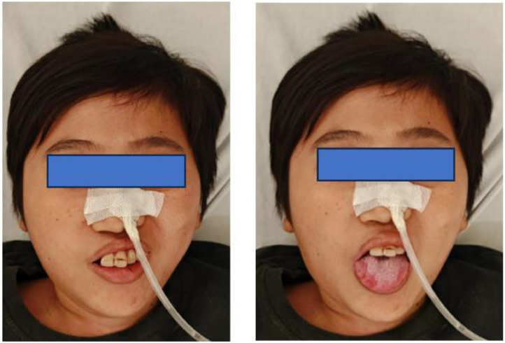
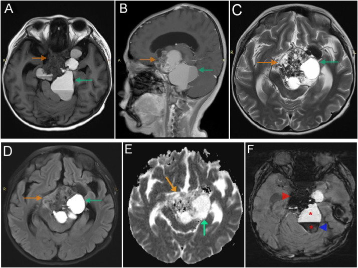
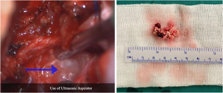
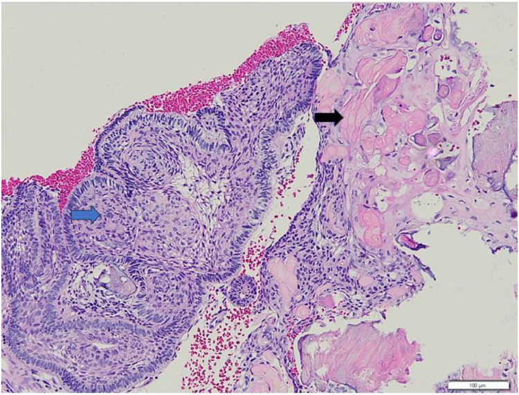
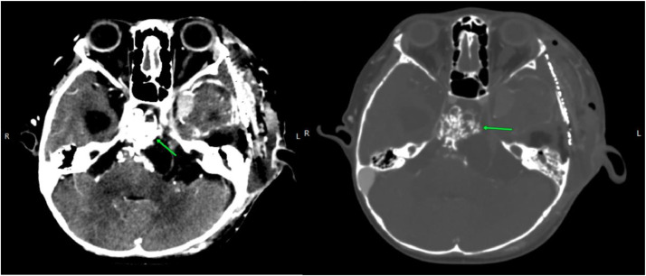
Craniopharyngiomas are rare, slow growing tumors arising along the craniopharyngeal duct. The incidence of craniopharyngioma was 0.13 per 100,000 persons per year. Intratumoral hemorrhage is a serious complication that can mimic pituitary tumor apoplexy, a life-threatening condition. In this report, We presented a case of a 20-year-old male with 3 days of decreased consciousness, with a history of worsening headaches, vomiting, blurry vision, bitemporal hemianopia, and right-sided limb weakness. MRI findings revealed a mixed cystic and solid suprasellar mass with blooming artifacts and fluid-fluid levels on SWI strongly suggest craniopharyngiomas with intratumoral hemorrhage. Trans-petrosal surgery was performed, the lesion appeared intraoperatively as a firm, elastic mass with a hemorrhagic component. Further histopathological testing confirmed the diagnosis of craniopharyngioma with adamantinomatous subtype, which typically occurs in children and adults over 45. While in this patient's diagnosis at age 20 falls outside the usual range. This study highlights the possibility of adamantinomatous craniopharyngioma occurring in young adulthood and the role of imaging in diagnosing craniopharyngioma, including its detailed characteristics and the presence of intratumoral hemorrhage for early management and better patient outcome.

Keywords: Adamantinomatous; Craniopharyngiomas; Intrasellar tumours; Intratumoral hemorrhage; Suprasellar.


Figures

Fig 1
Fig. 1
Clinical image shows facial asymmetry and mouth turned to the left side. There was confirmed left cranial nerve VII paresis.
Fig 2
Fig. 2
Head MRI shows a mixed mass: Solid components (orange arrow) give hypointense on T1 (A), inhomogeneous enhancement (B), heterogeneous isointense on T2 and FLAIR (C and D), no diffusion restriction (E), blooming artifacts on SWI (F, red arrowhead). Cystic components (green arrow) give hyperintense on T1, T2, and FLAIR (A-C-D), no enhancement (B), no diffusion restriction (E), with some showing blooming artifacts on SWI (F, blue arrowhead) with fluid-fluid levels (red star).
Fig 3
Fig. 3
Intraoperative examination revealed segmental dissection of the tumor mass and the component of blood (blue arrow).
Fig 4
Fig. 4
Microscopic examination reveals epithelial tumor cells exhibiting hyperplastic growth. The tumor periphery demonstrates the palisading architecture of the tumor cells, and the nuclei tend to be monomorphic with smooth chromatin and regular nuclear membrane. The central area displays stellate reticulum cells (blue arrow). Wet keratin is also seen (black arrow). (Magnification 100x, H&E stain).
Fig 5
Fig. 5
Axial post contrast head CT showed a enhanced residual solid mass with calcification (green arrow) in the suprasellar region.

Similar articles

References

    1. Muller HL, Merchant TE, Warmuth-Metz M, Martinez-Barbera JP, Puget S. Craniopharyngioma. Nat Rev Dis Primers. 2019;5:75. doi: 10.1038/s41572-019-0125-9. - DOI - PubMed
    1. Bunin GR, Surawicz TS, Witman PA, Preston-Martin S, Davis F, Bruner JM. The descriptive epidemiology of craniopharyngioma. J Neurosurg. 1998;89:547–551. doi: 10.3171/jns.1998.89.4.0547. - DOI - PubMed
    1. Chen Y, Hu F, Wang J, Huang K, Liu W, Tan Y, et al. Clinical features of craniopharyngioma with tumoral hemorrhage: a retrospective case-controlled study. Front Surg. 2022;9 doi: 10.3389/fsurg.2022.845273. - DOI - PMC - PubMed
    1. Prabhu VC, Brown HG. The pathogenesis of craniopharyngiomas. Childs Nerv Syst. 2005;21(8-9):622–627. doi: 10.1007/s00381-005-1190-9. - DOI - PubMed
    1. Yaşargil MG, Curcic M, Kis M, Siegenthaler G, Teddy PJ, Roth P. Total removal of craniopharyngiomas. Approaches and long-term results in 144 patients. J. Neurosurg. 1990;73:3–11. doi: 10.3171/jns.1990.73.1.0003. [PubMed] [Google Scholar] - DOI - PubMed

Publication types

LinkOut - more resources