. 2024 Dec 3;148(1):78.
doi: 10.1007/s00401-024-02824-9.

Identification of isoAsp7-Aβ as a major Aβ variant in Alzheimer's disease, dementia with Lewy bodies and vascular dementia

Affiliations

Identification of isoAsp7-Aβ as a major Aβ variant in Alzheimer's disease, dementia with Lewy bodies and vascular dementia

Sarah Schrempel et al. Acta Neuropathol. .

Abstract

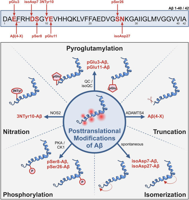
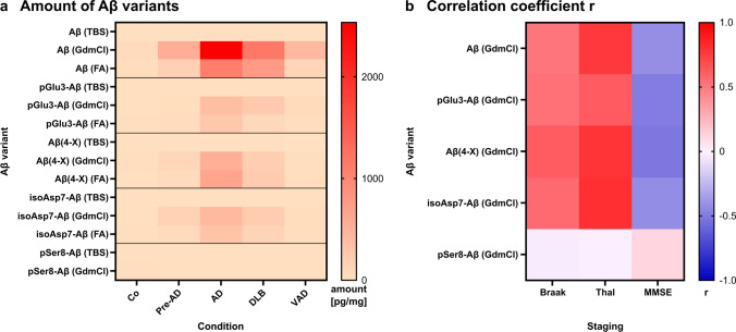
The formation of amyloid-β (Aβ) aggregates in brain is a neuropathological hallmark of Alzheimer's disease (AD). However, there is mounting evidence that Aβ also plays a pathogenic role in other types of dementia and that specific post-translational Aβ modifications contribute to its pathogenic profile. The objective of this study was to test the hypothesis that distinct types of dementia are characterized by specific patterns of post-translationally modified Aβ variants. We conducted a comparative analysis and quantified Aβ as well as Aβ with pyroglutamate (pGlu3-Aβ and pGlu11-Aβ), N-truncation (Aβ(4-X)), isoaspartate racemization (isoAsp7-Aβ and isoAsp27-Aβ), phosphorylation (pSer8-Aβ and pSer26-Aβ) or nitration (3NTyr10-Aβ) modification in post mortem human brain tissue from non-demented control subjects in comparison to tissue classified as pre-symptomatic AD (Pre-AD), AD, dementia with Lewy bodies and vascular dementia. Aβ modification-specific immunohistochemical labelings of brain sections from the posterior superior temporal gyrus were examined by machine learning-based segmentation protocols and immunoassay analyses in brain tissue after sequential Aβ extraction were carried out. Our findings revealed that AD cases displayed the highest concentrations of all Aβ variants followed by dementia with Lewy bodies, Pre-AD, vascular dementia and non-demented controls. With both analytical methods, we identified the isoAsp7-Aβ variant as a highly abundant Aβ form in all clinical conditions, followed by Aβ(4-X), pGlu3-Aβ, pGlu11-Aβ and pSer8-Aβ. These Aβ variants were detected in distinct plaque types of compact, coarse-grained, cored and diffuse morphologies and, with varying frequencies, in cerebral blood vessels. The 3NTyr10-Aβ, pSer26-Aβ and isoAsp27-Aβ variants were not found to be present in Aβ plaques but were detected intraneuronally. There was a strong positive correlation between isoAsp7-Aβ and Thal phase and a moderate negative correlation between isoAsp7-Aβ and performance on the Mini Mental State Examination. Furthermore, the abundance of all Aβ variants was highest in APOE 3/4 carriers. In aggregation assays, the isoAsp7-Aβ, pGlu3-Aβ and pGlu11-Aβ variants showed instant fibril formation without lag phase, whereas Aβ(4-X), pSer26-Aβ and isoAsp27-Aβ did not form fibrils. We conclude that targeting Aβ post-translational modifications, and in particular the highly abundant isoAsp7-Aβ variant, might be considered for diagnostic and therapeutic approaches in different types of dementia. Hence, our findings might have implications for current antibody-based therapies of AD.

Keywords: Abeta; Alzheimer’s disease; Amyloid-β; Automated histology quantification; Dementia with Lewy bodies; Mini Mental State Examination; Post-translational modifications; Vascular dementia.


Conflict of interest statement

Declarations. Conflict of interest: The authors declare no competing interests. Consent for publication: All the authors have approved publication.

Figures

Fig. 1
Fig. 1
Schematic representation of the Aβ peptide variants investigated in the present study in brain tissue of deceased patients categorized in Pre-AD, AD, DLB, VAD and control subjects. Together, eight different Aβ variants from five groups of post-translational modifications were analyzed side-by-side in a comprehensive manner by immunohistochemical and biochemical methods. Aβ structure from [25]. Created with BioRender.com
Fig. 2
Fig. 2
Schematic presentation of the workflow for the immunohistochemical (top) and biochemical (bottom) analyses of Aβ peptide variants in human brain tissue from Pre-AD, AD, DLB, VAD and control subjects. The immunohistochemically labeled brain slices were digitized with an Axioscan slide scanner and subjected to plaque load quantification by machine learning-based segmentation protocols. For biochemical analyses, Aβ peptides were extracted from unfixed human brain tissue by sequential centrifugation of Tris-buffered saline (TBS), guanidinium chloride (GdmCl) and formic acid (FA) dilutions followed by quantification by immunoassays using specific monoclonal antibodies. Created with BioRender.com
Fig. 3
Fig. 3
Representative examples of immunohistochemical labeling of human cortical brain tissue from control cases and different clinical conditions using antibodies to detect specific post-translational Aβ modifications as indicated (left). Respective quantifications of Aβ plaque load as a percentage of brain area covered by plaques is presented (right). Note that the Y-axes differ between the individual Aβ variants. Differences between clinical groups are statistically significant at *p < 0.05; **p < 0.01; ***p < 0.001. Medians are indicated by horizontal lines
Fig. 4
Fig. 4
a Subgroup analyses of the abundance of Aβ variants by APOE genotype. Note the high plaque load for all Aβ variants in APOE 3/4 carriers and the particularly high abundance of isoAsp7-Aβ compared to the other Aβ PTMs in this subgroup. b Pearson correlation analyses between the immunohistochemically quantified plaque load for individual Aβ variants and the neuropathological Braak Tau stages (column 1) and Thal amyloid phases (column 2). In addition, correlations between the respective abundance of Aβ variants and the MMSE scores (column 3) are presented. Pearson correlation was considered as moderate with correlation coefficients of 0.40 ≤ r < 0.70 (highlighted in light grey) and as strong with 0.70 ≤ r < 0.90 (highlighted in grey). Note the moderate and strong correlations of isoAsp7-Aβ with Braak Tau stages, Thal amyloid phases and MMSE scores. The individual cases relate to the clinical condition as follows: black—Co; grey—Pre-AD; red—AD; blue—DLB; green—VAD
Fig. 5
Fig. 5
Heat maps for the plaque load of Aβ variants and the correlation coefficient r of the Pearson correlation analyses. a Immunohistochemically quantified plaque load of Aβ variants is depicted for the different conditions. b For all correlations between the plaque load of Aβ variants and Braak and Thal staging and MMSE score, the correlation coefficient r as a measure for the quality of the correlation is presented. Note that the typical correlation pattern is not observed for pSer8-Aβ
Fig. 6
Fig. 6
a Association of Aβ, pGlu3-Aβ, Aβ(4-X), isoAsp7-Aβ, pSer8-Aβ and pGlu11-Aβ variants with amyloid plaques of compact, coarse-grained, cored and diffuse morphologies as well as with cerebral vessels. The images on the top show labeling of Aβ. The quantifications below indicate a high abundance for most Aβ variants in all plaque types, in particular in AD and DLB. In blood vessels, pSer8-Aβ and pGlu11-Aβ variants were particularly abundant in all clinical conditions, whereas pGlu3-Aβ and isoAsp7-Aβ were present in a smaller subset of patients in all clinical conditions. b Aggregation curves of time-dependent fibril formation of the Aβ variants. Note the instant fibril formation of pGlu3-Aβ(1–40). As compared to unmodified Aβ(1–40), the variants pGlu3-Aβ(1–40), isoAsp7-Aβ(1–40), pSer8-Aβ(1–40) and 3NTyr10-Aβ(1–40) showed shorter lag phases, indicating more rapid formation of β-sheet containing aggregates. We did not observe significant fibril formation of Aβ(4–40), pSer26-Aβ(1–40) and isoAsp27-Aβ(1–40) variants. tlag—lag phase, t1/2—half maximum ThT fluorescence intensity time. c Electron microscopic images of fibrils derived from the respective Aβ variants
Fig. 7
Fig. 7
Quantification of Aβ variants by immunoassays. Note that the Y-axes differ for the individual Aβ variants and for TBS, GdmCl and FA fractions. Differences between clinical groups are statistically significant at *p < 0.05; **p < 0.01; ***p < 0.001. Medians are indicated by horizontal lines
Fig. 8
Fig. 8
a Subgroup analyses of the abundance of Aβ variants by APOE genotype in GdmCl fractions. Note the high quantity of Aβ in APOE 3/3 and APOE 3/4 carriers. b Pearson correlation analyses between the load for individual Aβ variants quantified by immunoassays in the GdmCl fractions and the neuropathological Braak Tau stages (column 1) and Thal amyloid phases (column 2). In addition, correlations between the abundance of Aβ variants and the MMSE scores (column 3) are presented. Pearson correlation was considered as moderate with correlation coefficients of 0.40 ≤ r < 0.70 (highlighted in light grey) and as strong with 0.70 ≤ r < 0.90 (highlighted in grey). The individual cases relate to the clinical condition as follows: black—Co; grey—Pre-AD; red—AD; blue—DLB; green – VAD
Fig. 9
Fig. 9
Heat maps for the amount of Aβ variants quantified in the TBS, GdmCl and FA fractions and the correlation coefficient r of the Pearson correlation analyses. a Aβ variants quantified by immunoassays are depicted for the different clinical conditions. b For all correlations between the Aβ concentrations and Braak and Thal staging and MMSE score, the correlation coefficient r as a measure for the quality of the correlation is presented. Note that the typical correlation pattern is not observed for pSer8-Aβ

Similar articles

Cited by

References

    1. Abramov E, Dolev I, Fogel H, Ciccotosto GD, Ruff E, Slutsky I (2009) Amyloid-beta as a positive endogenous regulator of release probability at hippocampal synapses. Nat Neurosci 12:1567–1576. 10.1038/nn.2433 - PubMed
    1. Alexandru A, Jagla W, Graubner S, Becker A, Bäuscher C, Kohlmann S et al (2011) Selective hippocampal neurodegeneration in transgenic mice expressing small amounts of truncated Aβ is induced by pyroglutamate-Aβ formation. J Neurosci 31:12790–12801. 10.1523/JNEUROSCI.1794-11.2011 - PMC - PubMed
    1. Antzutkin ON, Leapman RD, Balbach JJ, Tycko R (2002) Supramolecular structural constraints on Alzheimer’s beta-amyloid fibrils from electron microscopy and solid-state nuclear magnetic resonance. Biochemistry 41:15436–15450. 10.1021/bi0204185 - PubMed
    1. Arezoumandan S, Cousins KAQ, Ohm DT, Lowe M, Chen M, Gee J et al (2024) Tau maturation in the clinicopathological spectrum of Lewy body and Alzheimer’s disease. Ann Clin Transl Neurol 11:673–685. 10.1002/acn3.51988 - PMC - PubMed
    1. Arima K, Uéda K, Sunohara N, Arakawa K, Hirai S, Nakamura M et al (1998) NACP/alpha-synuclein immunoreactivity in fibrillary components of neuronal and oligodendroglial cytoplasmic inclusions in the pontine nuclei in multiple system atrophy. Acta Neuropathol 96:439–444. 10.1007/s004010050917 - PubMed

Publication types