. 2024 Dec 10;121(50):e2404877121.
doi: 10.1073/pnas.2404877121. Epub 2024 Dec 3.

TRPC6 is a mechanosensitive channel essential for ultrasound neuromodulation in the mammalian brain

Affiliations

TRPC6 is a mechanosensitive channel essential for ultrasound neuromodulation in the mammalian brain

Yumi Matsushita et al. Proc Natl Acad Sci U S A. .

Abstract

Ultrasound neuromodulation has become an innovative technology that enables noninvasive intervention in mammalian brain circuits with high spatiotemporal precision. Despite the expanding utility of ultrasound neuromodulation in the neuroscience research field and clinical applications, the molecular and cellular mechanisms by which ultrasound impacts neural activity in the brain are still largely unknown. Here, we report that transient receptor potential canonical 6 (TRPC6), a mechanosensitive nonselective cation channel, is essential for ultrasound neuromodulation of mammalian neurons in vitro and in vivo. We first demonstrated that ultrasound irradiation elicited rapid and robust Ca2+ transients mediated via extracellular Ca2+ influx in cultured mouse cortical and hippocampal neurons. Ultrasound-induced neuronal responses were massively diminished by blocking either the generation of action potential or synaptic transmission. Importantly, both pharmacological inhibition and genetic deficiency of TRPC6 almost completely abolished neuronal responses to ultrasound. Furthermore, we found that intracerebroventricular administration of a TRPC6 blocker significantly attenuated the number of neuronal firings in the cerebral cortex evoked by transcranial ultrasound irradiation in mice. Our findings indicate that TRPC6 is an indispensable molecule of ultrasound neuromodulation in intact mammalian brains, providing fundamental understanding of biophysical molecular mechanisms of ultrasound neuromodulation as well as insight into its future feasibility in neuroscience and translational research in humans.

Keywords: TRPC6; cortical neuron; mechanosensitive channels; neuromodulation; ultrasound.


Conflict of interest statement

Competing interests statement:The authors declare no competing interest.

Figures
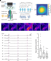
Fig. 1.
Fig. 1.
Ultrasound irradiation induces Ca2+ transients in cultured cortical neurons. (A) A scheme of in vitro experimental setup to monitor ultrasound-induced neuronal response. (B) An ultrasound burst pulse sequence used for in vitro experiments. PRF: Pulse repetition frequency, DC: Duty cycle. (C) Pressure distribution of stimulating ultrasound measured inside free water. (D) Representative sequential images depicting GCaMP6s fluorescence in neuronal cells during pre-, post-, and peristimulation phases induced by ultrasound irradiation. The scale bar demonstrates minimum to maximum values normalized to a global max. (E) Representative traces of fluorescence calcium imaging demonstrating pulsed ultrasound irradiation induced rapid Ca2+ transients in individual neurons. Ultrasound stimulation at an intensity of 190 mW/cm2 and duration of 500 msec is applied to neurons at the time of US1, US2, and US3. (F) The amplitudes of Ca2+ transients in response to sequential ultrasound stimulation at US1, US2, and US3 are quantified by area under the curve (AU). Bar graph values show mean ± SEM (n = 11, P = 0.1349, one-way ANOVA).
Fig. 2.
Fig. 2.
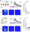
Ultrasound-mediated Ca2+ transients depend on extracellular Ca2+ influx and network activity. (A) A scheme of strategies to investigate the impact of Ca2+ influx from outside of the cell on ultrasound-induced neuronal response. Cd2+ (100 µM) blocks Ca2+-permeable ion channels. (B) Averaged traces of neuronal Ca2+ transients against ultrasound irradiation under the condition of control (black), Ca2+ free (red), or the presence of Cd2+(blue). Data from seven, nine, and eight independent experiments for control, Ca2+ free, and Cd2+ are plotted as mean ± SEM, respectively. The arrowhead indicates the time of ultrasound stimulation. (C) Heatmap demonstration of normalized fluorescence intensity (ΔF/F0) of GCaMP6s in neurons under the condition of control, Ca2+ free, or the presence of Cd2+. Data from 60 cells in one experiment are plotted. Arrowheads indicate the time of ultrasound stimulation. (D) The amplitudes of Ca2+ transients in response to ultrasound stimulation are quantified as the area under the curve (AU). Bar graph values show mean ± SEM. **P < 0.01 (one-way ANOVA followed by the Dunnett post hoc test). (E) A scheme of strategies to investigate the impact of the generation of action potentials and functional network activity on ultrasound-induced neuronal response. TTX (1 µM) is used to prevent the generation of action potentials and a mixture of CNQX (10 µM) and AP5 (50 µM) is used to block synaptic transmission. (F) Averaged traces of neuronal Ca2+ transients against ultrasound irradiation under the condition of control (black), TTX-treated (orange), or CNQX+AP5-treated (purple) neurons. Data from six, six, and eight independent experiments for control, TTX, and CNQX+AP5 were plotted as mean ± SEM, respectively. (G) Heatmap analysis of neuronal responses under the condition of control, TTX-treated, or CNQX+AP5-treated neurons. Data from 60 cells in one experiment were plotted. (H) Bar graph values show mean ± SEM. *P < 0.05 (one-way ANOVA followed by the Dunnett post hoc test).
Fig. 3.
Fig. 3.
Pharmacological investigation of the involvement of mechanosensitive channels in neuronal responses to ultrasound stimulation. (A) A schematic diagram of mechanosensitive receptors and effects of antagonists. RuR (RuR, 5 µM) was used to block TRPVs channel and GsMT×4 (10 µM) was used to prevent gating Piezo1/2, TRPC1, and TRPC6. (B) Averaged traces of neuronal Ca2+ transients against ultrasound irradiation under the condition of control (black), RuR-treated (red), or GsMT×4-treated (blue) neurons. Data from five, six, and six independent experiments for control, RuR, and GsMTx4 are described as mean ± SEM, respectively. The arrowhead indicates the time of ultrasound stimulation. (C) Heatmap demonstration of normalized fluorescence intensity (ΔF/F0) of GCaMP6s in neurons under the condition of control, or the presence of RuR or GsMT×4. Arrowheads indicate the time of ultrasound stimulation. Data from 60 cells in an experiment are plotted. Arrowheads indicate the time of ultrasound stimulation. (D) The amplitude of Ca2+ transients in responses to ultrasound stimulation is quantified as area under the curve (AU). Bar graph values show mean ± SEM. *P < 0.05 (one-way ANOVA followed by the Dunnett post hoc test). (E) A scheme of the effects of selective antagonists on Piezo1 or TRPC6. Dooku1 (10 µM) and BI-749327 (1 µM) were used to inhibit Piezo1 and TRPC6 activation, respectively. (F) Averaged traces of neuronal Ca2+ transients against ultrasound irradiation under the condition of control (black), or the presence of Dooku1 (purple) or BI-749327 (light blue). Fourteen, thirteen, and twelve independent experiments for control, Dooku1, and BI-749327, respectively, were performed. Traces show mean ± SEM. (G) Heatmap analysis of neuronal responses under the condition of control, Dooku1 treated, or BI-749327 treated neurons. (H) Bar graph values of area under the curve in each condition show mean ± SEM. **P < 0.01 (one-way ANOVA followed by the Dunnett post hoc test).
Fig. 4.
Fig. 4.
TRPC6-KO neurons lack the response to ultrasound irradiation that can be rescued by overexpression of TRPC6. (A) RT-PCR validation of TRPC6 mRNA expression in total RNA extracted from cultured neurons of wild-type (WT), TRPC6 deficient (KO), and TRPC6-KO overexpressing mouse TRPC6-HA (OE) mice. Representative gel electrophoresis images from 2 samples each shows PCR products targeting TRPC6 (327 bp). (B) Averaged neuronal responses to ultrasound irradiation of WT (black), TRPC6-KO (orange), or TRPC6-KO overexpressing mouse TRPC6-HA (TRPC6-KO w/ mTRPC6) (blue) neurons. Twelve, eleven, and twelve independent experiments for WT, TRPC6-KO, and TRPC6-KO w/ mTRPC6 neurons, respectively, were performed. The arrowhead indicates the time of ultrasound stimulation. Traces show mean ± SEM. (C) Heatmap demonstration of normalized fluorescence intensity (ΔF/F0) of GCaMP6s in WT, TRPC6-KO, and TRPC6-KO w/ mTRPC6. Arrowheads indicate the time of ultrasound stimulation. Data from 60 cells in two experiments are plotted. Arrowheads indicate the time of ultrasound stimulation. (D) Bar graph values of area under the curve in each condition show mean ± SEM. **P < 0.01 (one-way ANOVA followed by the Dunnett post hoc test).
Fig. 5.
Fig. 5.
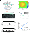
TRPC6 mediates ultrasound-induced neuronal activation in vivo. (A) An ultrasound burst pulse sequence used for in vivo experiments. (B) Pressure distribution of stimulating ultrasound measured inside free water. (C and D) Schematic diagrams of in vivo multiunit recordings with a TRPC6 blocker, BI-749327. (C) Ultrasound was transcranially irradiated to the cerebral cortex of mice, and a tungsten microelectrode for recordings was inserted through a cranial window. A white arrowhead indicates a representative recording site labeled with an electrical lesion after recordings. (D) BI-749327 was intracerebroventricularly administered 30 min before post recordings. (E and F) Representative in vivo multiunit recordings around US irradiations before (E) and after (F) BI-749327 administration. Top: high-pass filtered extracellular voltage traces; Bottom: peristimulus time histograms (PSTHs). Orange patches represent ultrasound irradiation. Dashed pink, red, and blue lines in each PSTH represent global significance levels, local significance levels, and mean counts calculated with 1,000 randomly generated datasets of timestamps of population neural activities with time jittering, respectively. The significance of each recording was determined by whether PSTH goes above or down across the global significant levels. (G) Fraction of significantly activated population responses by US irradiations of several intensities. Significance on each stimulus intensity was determined by the Χ2 test on a two-by-two (significance × control or BI-749327) frequency table. n = 73, 80 sessions from three mice for pre- and postrecordings, respectively. *P < 0.05.
Fig. 6.
Fig. 6.
A hypothetical model for the mechanism of ultrasound neuromodulation via TRPC6 activation proposed in the present study. Ultrasound irradiation potentially distorts the cell membrane by acoustic pressure, which triggers the opening of a mechanosensitive TRPC6 channel followed by membrane depolarization in cultured cortical neurons. The generation of action potentials may be induced by the opening of VGSC following the TRPC6-mediated depolarization, which may further elicit extracellular Ca2+ influx via voltage-gated calcium channels, resulting in excitation of the neuronal network.

References

    1. Temel Y., Jahanshahi A., Neuroscience treating brain disorders with neuromodulation. Science 347, 1418–1419 (2015). - PubMed
    1. Mosilhy E. A., et al. , Non-invasive transcranial brain modulation for neurological disorders treatment: A narrative review. Life Sci. 307, 120869 (2022). - PubMed
    1. Denison T., Morrell M. J., Neuromodulation in 2035: The neurology future forecasting series. Neurology 98, 65–72 (2022). - PMC - PubMed
    1. Darmani G., et al. , Non-invasive transcranial ultrasound stimulation for neuromodulation. Clin. Neurophysiol. 135, 51–73 (2022). - PubMed
    1. Song M., Zhang M., He S., Li L., Hu H., Ultrasonic neuromodulation mediated by mechanosensitive ion channels: Current and future. Front. Neurosci. 17, 1232308 (2023). - PMC - PubMed

LinkOut - more resources