. 2025 Feb 25;9(4):844-855.
doi: 10.1182/bloodadvances.2024013446.

TIFAB modulates metabolic pathways in KMT2A::MLLT3-induced AML through HNF4A

Affiliations

TIFAB modulates metabolic pathways in KMT2A::MLLT3-induced AML through HNF4A

Yang Wang et al. Blood Adv. .

Abstract

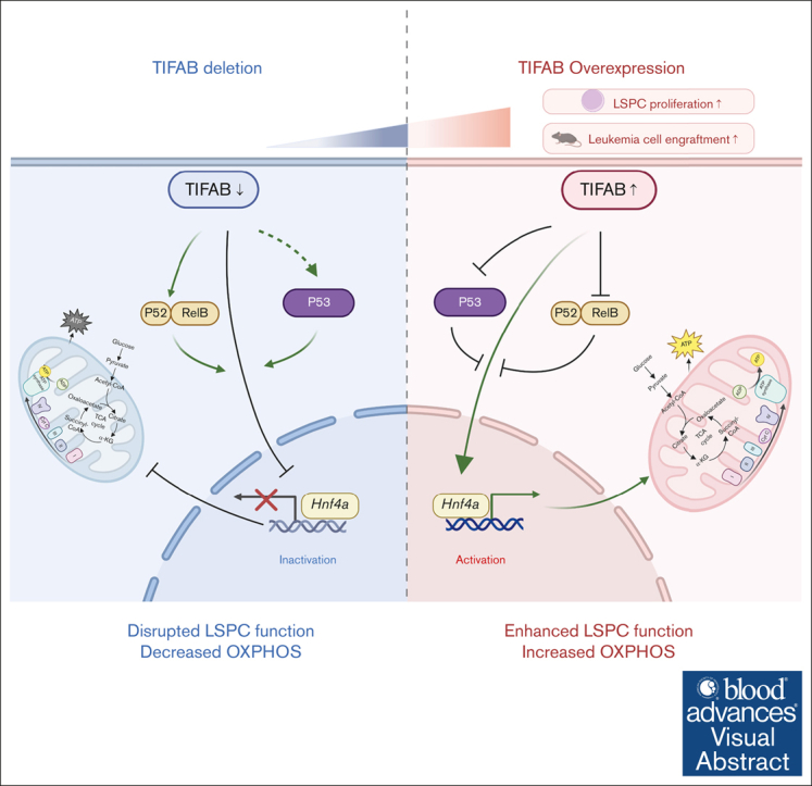
Tumor necrosis factor (TNF) receptor-associated factor (TRAF)-interacting protein with forkhead-associated domain B (TIFAB), an inhibitor of NF-κB signaling, plays critical roles in hematopoiesis, myelodysplastic neoplasms, and leukemia. We previously demonstrated that Tifab enhances KMT2A::MLLT3-driven acute myeloid leukemia (AML) by either upregulating Hoxa9 or through ubiquitin-specific peptidase 15-mediated downregulation of p53 signaling. In this study, we show that Tifab deletion in KMT2A::MLLT3-induced AML impairs leukemia stem/progenitor cell (LSPC) engraftment, glucose uptake, and mitochondrial function. Gene set enrichment analysis reveals that Tifab deletion downregulates MYC, HOXA9/MEIS1, mTORC1 signaling, and genes involved in glycolysis and oxidative phosphorylation. By comparing genes upregulated in TIFAB-overexpressing LSPCs with those downregulated upon Tifab deletion, we identify hepatocyte nuclear factor 4 alpha (Hnf4a) as a key TIFAB target, regulated through the inhibition of NF-κB component RelB, which suppresses Hnf4a in leukemia cells. HNF4A, a nuclear receptor involved in organ development, metabolism, and tumorigenesis, rescues the metabolic defects caused by Tifab deletion and enhances leukemia cell engraftment. Conversely, Hnf4a knockdown attenuates TIFAB-mediated enhancement of LSPC function. These findings highlight the critical role of the TIFAB-HNF4A axis in KMT2A::MLLT3-induced AML and uncover a novel regulator in leukemia biology.


Conflict of interest statement

Conflict-of-interest disclosure: D.T.S. serves on the scientific advisory board at Kurome Therapeutics; is a consultant for and/or received funding from Kurome Therapeutics, Captor Therapeutics, Treeline Biosciences, and Tolero Therapeutics; and has equity in Kurome Therapeutics. The remaining authors declare no competing financial interests.

Figures

None
Graphical abstract
Figure 1.
Figure 1.
TIFAB overexpression enhances LSPC OXPHOS activity. (A) GSEA of RNA-seq data comparing vector control (Con) and TIFAB-overexpressing (TIFAB OE) LSPCs isolated from leukemic mice transplanted with either empty vector or TIFAB OE KMT2A::MLLT3 leukemia cells, indicating upregulation of OXPHOS with TIFAB overexpression. (B-C) OCR (Mito Stress test) (B) and ECAR (Glycolysis Stress test) (C) in Con and TIFAB OE LSPCs (n = 5). (D-F) Mitochondrial mass (n = 6) (D), mitochondrial membrane potential (n = 7) (E), and ROS levels (n = 7) (F) were assessed using MitoTracker, tetramethylrhodamine ethyl ester (TMRE), and CellROX, respectively, in Con and TIFAB OE LSPCs. (G) Fractional enrichment of 13C6-labeled intermediate metabolites in Con and TIFAB OE LSPCs, measured by GC/MS (n = 3). ∗P < .05; ∗∗P < .01. Tests used in panels D-G, Mann-Whitney U test. (H) Western blot analysis of key components of the ETC complexes I, II, III, IV, and V, as well as HNF4A and TIFAB in Con and TIFAB OE LSPCs (n = 3). ACTIN served as a loading control. FDR, false discovery rate; NES, normalized enrichment score.
Figure 2.
Figure 2.
Tifab deletion impairs cellular functions of LSPCs. (A) Cell counting of WT and Tifab KO LSPCs. Ten thousand CD11b+cKit+ leukemia cells were plated and counted every other day for 4 days. (B) Percentage of Ki67+ LSPCs in mice that received transplant with WT or Tifab KO KMT2A::MLLT3 leukemia cells. (C) Percentage of caspase-3–positive LSPCs in WT and Tifab KO LSPCs. (D) Representative flow cytometry profiles of mice engrafted with WT or Tifab KO LSPCs, assessed 8 weeks after transplantation. (E) Survival curve of sublethally irradiated recipient mice receiving 20 000 sorted LSPCs from WT or Tifab KO KMT2A::MLLT3 leukemic mice (WT, n = 7; Tifab KO, n = 8). (F-G) OCR (F) and ECAR (G) in WT and Tifab KO LSPCs (n = 5). (H) Fractional enrichment of 13C6-labeled intermediate metabolites in Con and Tifab KO LSPCs, measured by GC/MS (n = 3). (I) Western blot analysis of key components of ETC complexes I, II, III, IV, and V, along with TIFAB, in WT and Tifab KO LSPCs (n = 3). ACTIN was used as a loading control. ∗P < .05; ∗∗P < .01. Statistical test used in panels A-C and H was Mann-Whitney U test; for panel E, log-rank test.
Figure 3.
Figure 3.
Tifab deletion disrupts signaling pathways regulating LSPC function. (A-F) GSEA of RNA-seq data comparing WT and Tifab KO LSPCs. Tifab KO LSPCs exhibit downregulation of gene sets associated with OXPHOS (A), Myc targets (B), HOXA9 and MEIS1 signaling (C), MTORC1 signaling (D), KMT2A::MLLT3 fusion targets (E), and glycolysis (F), compared with WT LSPCs. (G) Quantitative polymerase chain reaction analysis of glycolysis-related enzyme expression in WT and Tifab KO LSPCs. Representative results from 3 independent experiments, each performed with 3 replicates. (H-I) Glucose uptake in WT and Tifab KO (n = 7) (H) or Con and TIFAB OE (n = 16) LSPCs (I). ∗P < .05; ∗∗P < .01. In panel G, Mann-Whitney U test was used; in panels H-I, Student t test was used. FDR, false discovery rate; NES, normalized enrichment score.
Figure 4.
Figure 4.
Hnf4a is a downstream target of TIFAB, negatively regulated by NF-κB. (A) Venn Diagram illustrating genes upregulated or downregulated in TIFAB OE KMT2A::MLLT3 LSPCs (compared with vector-expressed, GSE178853) and Tifab KO LSPCs (compared with WT). Among these genes, 117 were upregulated in TIFAB OE LSPCs while downregulated in Tifab KO LSPCs, including Hnf4a. (B-C) Expression of HNF4A protein in vector- and KMT2A::MLLT3−transduced cKit+ WT BM cells (B) or WT and Tifab KO LSPCs (n = 3). ACTIN served as a loading control. (D-E) Hnf4a mRNA (D) or protein (E) levels in KMT2A::MLLT3 LSPCs expressing either vector, RelA, or RelB. ∗P < .05, Mann-Whitney U test. (F) Higher expression of HNF4A mRNA in patients with AML (TCGA database) correlates with worse survival. P = .0052, log-rank test.
Figure 5.
Figure 5.
HNF4A overexpression rescues Tifab deletion–induced LSPC dysfunction. (A-B) HNF4A overexpression increases LSPC proliferation (n = 5) (A) and enhances colony formation (n = 5) (B). (C) Glucose uptake assessed by NBDG incorporation in WT or Tifab KO LSPCs transfected with either vector- (Vec) or HNF4A-expressing viruses (n = 5). (D-F) Mitochondrial mass (n = 5) (D), mitochondrial membrane potential (n = 5) (E), and ROS levels (n = 10) (F) in WT or Tifab KO LSPCs transfected with either Vec- or HNF4A-expressing viruses, measured using MitoTracker, TMRE, and CellROX, respectively. (G-H) OCR (G) and ECAR (H) in WT or Tifab KO LSPCs transfected with either Vec- or HNF4A-expressing viruses (n = 5). (I) Western blot analysis of key components of ETC complexes I, II, III, IV, and V, as well as TIFAB and HNF4A, in WT and Tifab KO LSPCs transfected with either Vec- or HNF4A-expressing viruses (n = 3). ACTIN served as a loading control. (J) Proliferation of WT or Tifab KO LSPCs transfected with either Vec- or HNF4A-expressing viruses (n = 8). (K) Survival curve of sublethally irradiated recipient mice transplanted with Vec- or HNF4A-transduced WT or Tifab KO KMT3A::MLLT3 LSPCs (n = 8 in each group). ∗P < .05; ∗∗P < .01. Statistical tests used in panels A-B, Mann-Whitney U test; in panels C-F and J, 1-way analysis of variance and Tukey multiple comparisons test; in panel K, log-rank test.

References

    1. Tallman MS, Wang ES, Altman JK, et al. Acute myeloid leukemia, version 3.2019, NCCN Clinical Practice Guidelines in Oncology. J Natl Compr Canc Netw. 2019;17(6):721–749. - PubMed
    1. Khwaja A, Bjorkholm M, Gale RE, et al. Acute myeloid leukaemia. Nat Rev Dis Primers. 2016;2 - PubMed
    1. Pollyea DA, Jordan CT. Therapeutic targeting of acute myeloid leukemia stem cells. Blood. 2017;129(12):1627–1635. - PubMed
    1. Becker MW, Jordan CT. Leukemia stem cells in 2010: current understanding and future directions. Blood Rev. 2011;25(2):75–81. - PubMed
    1. Kreso A, Dick JE. Evolution of the cancer stem cell model. Cell Stem Cell. 2014;14(3):275–291. - PubMed

MeSH terms

Substances