. 2024 Dec 16;4(12):100910.
doi: 10.1016/j.crmeth.2024.100910. Epub 2024 Dec 2.

Enhancing immuno-oncology investigations through multidimensional decoding of tumor microenvironment with IOBR 2.0

Affiliations

Enhancing immuno-oncology investigations through multidimensional decoding of tumor microenvironment with IOBR 2.0

Dongqiang Zeng et al. Cell Rep Methods. .

Abstract

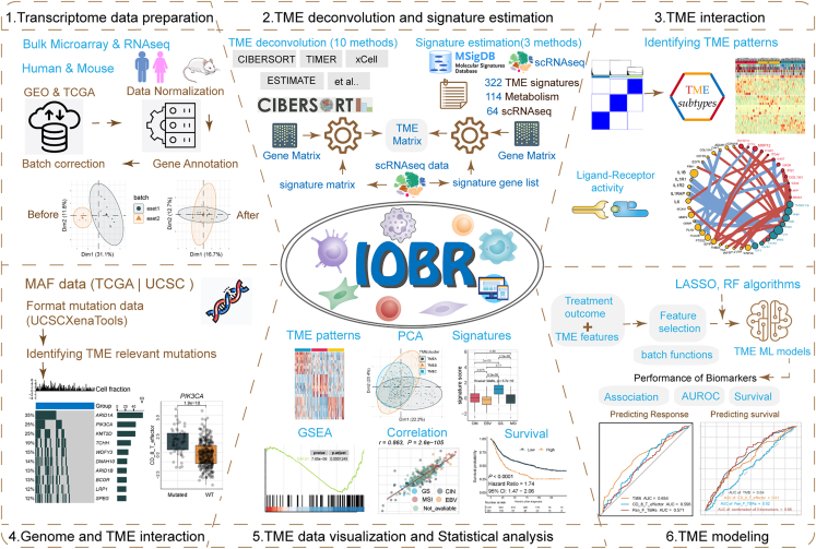
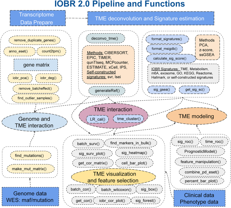
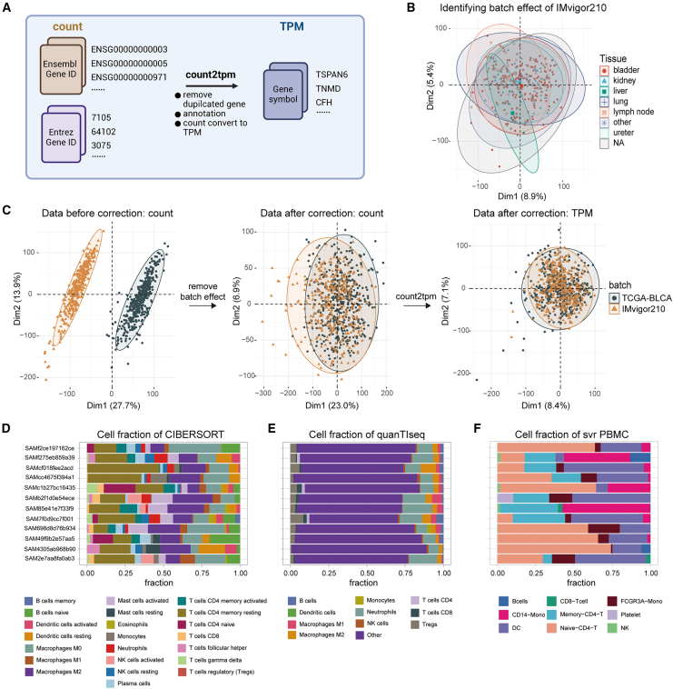
The use of large transcriptome datasets has greatly improved our understanding of the tumor microenvironment (TME) and helped develop precise immunotherapies. The growing application of multi-omics, single-cell RNA sequencing (scRNA-seq), and spatial transcriptome sequencing has led to many new insights, yet these findings still require clinical validation in large cohorts. To advance multi-omics integration in TME research, we have upgraded the Immuno-Oncology Biological Research (IOBR) package to IOBR 2.0, restructuring and standardizing its analytical workflow. IOBR 2.0 offers six modules for TME analysis based on multi-omics data, including data preprocessing, TME estimation, TME infiltration pattern identification, cellular interaction analysis, genome and TME interaction, and feature visualization, as well as modeling. Additionally, IOBR 2.0 enables constructing gene signatures and reference matrices from scRNA-seq data for TME deconvolution. The user-friendly pipeline provides comprehensive insights into tumor-immune interactions, and a detailed GitBook(https://iobr.github.io/book/) offers a complete manual and analysis guide for each module.

Keywords: CP: cancer biology; gene signatures; immunotherapy; multi-omics; single-cell data; tumor microenvironment; tumor-immune interaction; tumor-metabolism.


Conflict of interest statement

Declaration of interests The authors declare no competing interests.

Figures

None
Graphical abstract
Figure 1
Figure 1
The graphical scheme describing the workflow of the IOBR 2.0 IOBR 2.0 encompasses transcriptome data preparation, multiple deconvolution algorithms and signature estimation methods for microenvironment analysis, TME pattern identification, analysis of interactions between the genome and TME, batch visualization and statistical analysis, and TME modeling. TME, tumor microenvironment; MAF, mutation annotation format; PCA, principal-component analysis; GSEA, gene set enrichment analysis; RF, random forest; ML, machine learning; AUROC, area under the receiver operating characteristic curve.
Figure 2
Figure 2
IOBR 2.0 is composed of six analytic modules related to data preprocessing and tumor immune microenvironment The functionalities of these modules include (1) preprocessing of transcriptome data; (2) estimation of signature scores and identification of phenotype-relevant or user-constructed signatures, along with decoding the TME contexture; (3) identification of TME patterns and analysis of ligand-receptor interactions; (4) estimation of the specific mutation landscape associated with the signature of interest; (5) corresponding batch visualization and statistical analyses; and (6) model construction. WES, whole-exome sequencing.
Figure 3
Figure 3
Preprocessing of transcriptomic data and calculation of TME cell infiltration abundance (A) Flowchart illustrating the conversion of RNA-seq count data to TPM using the count2tpm function. (B) PCA scatterplot depicting the distribution of tissue types in the IMvigor210 dataset. (C) Comparison of data distribution before and after batch correction for IMvigor210 and TCGA-BLCA datasets. (D and E) Percentage bar plots displaying TME cell percentages based on CIBERSORT (D) and quanTIseq (E). (F) IOBR 2.0 utilizes cell-type-specific gene expression signatures generated from single-cell analysis to decode the TME landscape of the IMvigor210 cohort from bulk RNA-seq data. Each color represents a different cell type. NA, not available; TPM, transcripts per million.
Figure 4
Figure 4
IOBR 2.0 identifies TME patterns and analyzes differences in clinical features, phenotype, and TME components between TME clusters (A) Heatmap showing (blue = TME1; red = TME2) TME-infiltrating cell signature score of each patient in high (red) and low (blue) score groups. Rows of the heatmap represent TME-infiltrating cell signature expression (Z scores) calculated using the CIBERSORT algorithm via the deconvo_tme function. TME1 and TME2 were identified as distinct TME patterns using the tme_cluster function based on the TME-infiltrating cell signatures of CIBERSORT. (B) Kaplan-Meier curves comparing OS between TME1 and TME2. (C) Boxplots showing the expression of M1 and CD8+ T cell (Z scores), mutation burden per megabase (MB) and neoantigen burden per MB between TME1 and TME2. (D) Bar plot showing the clinical features and biomarkers, including immune phenotype, IC level, TC level, and BOR between TME1 and TME2. (E and F) The boxplot (E) and heatmap (F) delineate the metabolism signatures enrolled in IOBR 2.0, and identify signatures associated with the TME pattern. p value in (E) was calculated using two-sided Mann-Whitney U test. ∗p < 0.05; ∗∗p < 0.01; ∗∗∗p < 0.001; ∗∗∗∗p < 0.0001; ns, not significant compared to isotype group. IC level, immune cells level; TC level, tumor cells level; NA, not available; NE, not evaluable; CR, complete response; PR, partial response; SD, stable disease; PD, progressive disease; BOR, best of response.
Figure 5
Figure 5
Screening of immunotherapy prognostic features and construction of efficacy prediction models (A) The forest plots reveal 12 gene signatures correlated with OS in IMvigor210 cohort. (B) The correlation plot reflects the Spearman correlation between M1 macrophages and mutation burden per MB. (C) Kaplan-Meier curves comparing OS among four TME clusters (TME1–4). IOBR 2.0 identifies these four TME clusters based on 30 features closely related to immunotherapy prognosis. (D) Heatmap of immunotherapy prognosis-related signature scores derived from ssGSEA through IOBR 2.0 for the four TME clusters. (E) Kaplan-Meier curves comparing OS between high- and low-score group. The riskscore cutoff is set at the mean value. (F) The time-dependent ROC curves and AUC of riskscore (red), mutation burden per MB (gray), and the combined model (blue) predicting the clinical benefit for patients at 12 months. The combined model is constructed by integrating riskscore and mutation burden per MB using a Cox regression approach. ROC, receiver operating characteristic; ssGSEA, single-sample gene set enrichment analysis; OS, overall survival.
Figure 6
Figure 6
Genome and TME interaction module delineates mutations associated with TMEscore and the corresponding oncoplot (A) Boxplots displaying the mutations significantly associated with TMEscore, including TP53, FGFR3, ERBB3, and PIK3CA. The blue and yellow colors represent mutated and wild-type status, respectively. (B) Kaplan-Meier curves comparing OS between mutated and wild-type status of TP53. (C) Oncoprints depicting the genomic alteration landscapes in the context of high and low TMEscore. The numbers on the left green bars and on the right side collectively demonstrate the mutation frequency of each gene. WT, wild type; Mut, mutant.

References

    1. Robert C. A decade of immune-checkpoint inhibitors in cancer therapy. Nat. Commun. 2020;11:3801. doi: 10.1038/s41467-020-17670-y. - DOI - PMC - PubMed
    1. Morad G., Helmink B.A., Sharma P., Wargo J.A. Hallmarks of response, resistance, and toxicity to immune checkpoint blockade. Cell. 2021;184:5309–5337. doi: 10.1016/j.cell.2021.09.020. - DOI - PMC - PubMed
    1. de Visser K.E., Joyce J.A. The evolving tumor microenvironment: From cancer initiation to metastatic outgrowth. Cancer Cell. 2023;41:374–403. doi: 10.1016/j.ccell.2023.02.016. - DOI - PubMed
    1. Ye Z., Zeng D., Zhou R., Shi M., Liao W. Tumor Microenvironment Evaluation for Gastrointestinal Cancer in the Era of Immunotherapy and Machine Learning. Front. Immunol. 2022;13 doi: 10.3389/fimmu.2022.819807. - DOI - PMC - PubMed
    1. Zeng D., Wang M., Wu J., Lin S., Ye Z., Zhou R., Wang G., Wu J., Sun H., Bin J., et al. Immunosuppressive Microenvironment Revealed by Immune Cell Landscape in Pre-metastatic Liver of Colorectal Cancer. Front. Oncol. 2021;11 doi: 10.3389/fonc.2021.620688. - DOI - PMC - PubMed

LinkOut - more resources