. 2024 Dec 3;14(1):30150.
doi: 10.1038/s41598-024-81171-x.

Assessment of molecular modulation by multifrequency electromagnetic pulses to preferably eradicate tumorigenic cells

Affiliations

Assessment of molecular modulation by multifrequency electromagnetic pulses to preferably eradicate tumorigenic cells

Roberta Piredda et al. Sci Rep. .

Abstract

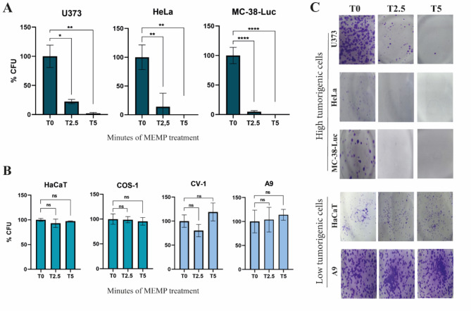
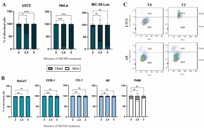
Physics methods of cancer therapy are extensively used in clinical practice, but they are invasive and often confront undesired side effects. A fully new equipment that allows sustained emission of intense and time-controlled non-ionizing multifrequency electromagnetic pulse (MEMP), has been applied to eukaryotic cells in culture. The equipment discriminates the overall electronegative charge of the cell cultures, and its subsequent proportional emission may thereby become higher and lethal to cancer cells of generally high metabolic activity. In contrast, low tumorigenic cells would be much less affected. We tested the specificity and efficacy of the equipment against a collection of (i) highly tumorigenic cells of human (glioblastoma, cervical carcinoma, and skin) and mouse (colon adenocarcinoma) origin; (ii) cell lines of much lower tumorigenicity (non-human primate kidney and mouse fibroblasts), and (iii) primary porcine macrophages lacking tumorigenicity. Time and intensity control of the MEMP allowed progressive decay of viability fairly correlating to cell tumorigenicity, which was provoked by a proportional alteration of the cytoplasmic membrane permeability, cell cycle arrest at G2, and general collapse of the actin cytoskeleton to the perinuclear region. Correspondingly, these effects drastically inhibited the proliferative capacity of the most tumorigenic cells in clonogenic assays. Moreover, MEMP suppressed in a dose-dependent manner the tumorigenicity of retrovirally transduced luciferase expressing colon adenocarcinoma cells in xenografted immune-competent mice, as determined by tumor growth in a bioluminescence imaging system. Our results support MEMP as an anti-cancer non-invasive physical treatment of substantial specificity for tumorigenic cells with promising therapeutic potential in oncology.


Conflict of interest statement

Declarations. Competing interests: The authors declare no competing interests.

Figures

Fig. 1
Fig. 1
Degree of inhibition of cell viability caused by MEMP as evaluated by an MTT assay. Quantitative determination of cell metabolic activity by MTT in non-treated controls and upon the indicated MEMP treatments. (A) Evaluation of cell viability in U373 MG, HeLa, and MC-38-Luc cancer cell lines treated from 0 to 5 min. (B) Evaluation of viability after MEMP treatment applied for 2.5 and 5 min to HaCaT, CV-1, COS-1, A9 cell lines, and PAM. Data are the mean with standard errors obtained from triplicates and were statistically analyzed by using a one-way ANOVA with a Dunnett’s test (*P < 0.05; **P < 0.01; ****P < 0.0001).
Fig. 2
Fig. 2
Differential inhibitory effect of MEMP treatments on cell lines assessed by colony forming ability. Control and modulated cell lines at the times indicated in the figure were seeded in triplicate at 103 to 104 cells onto P60 plates. The percentages of colonies formed in culture were calculated with respect to the number of colonies formed in the control (T0). (A) Percentage of colonies formed by U373, HeLa, and MC-38-Luc cells upon the MEMP treatments performed between 2.5 to 5 min. (B) Analysis of the inhibition of colony forming capacity caused by MEMP applied for 2.5 and 5 min to the HaCaT, COS-1, CV-1, and A9 cell lines. (C) Representative photographs showing the effect of MEMP treatment on colony formation by different cell lines. Colonies were fixed and stained with crystal violet two to three weeks after treatments. Data were statistically analyzed by using a one-way ANOVA with Dunnett’s test (*P < 0.05; **P < 0.01; ****P < 0.0001).
Fig. 3
Fig. 3
Determination of cell viability after MEMP treatment by cytometry. Cells modulated at times indicated in the figure were stained with the fixable Viability Dye Ghost Dye Red 780 (1 µg/mL) and fixed. Cells were then analyzed in a FACS Canto flow cytometer (BD Science) to determine the percentage of death and live cells. The figure shows representative data from three experiments on the percentages of live and dead cells after each MEMP modulation condition. Data were statistically analyzed by using a two-way ANOVA with Dunnett’s test (*P < 0.05; **P < 0.01; ****P < 0.0001). (A) Evaluation of MEMP effect on the viability of the U373, HeLa, and MC-38-Luc cancer cells. The experiment was repeated three times. (B) Determination of cell viability by FACS after the MEMP treatment in the HaCaT, CV-1, COS-1, A9 cell lines, and PAM. (C) Indicative photograph of dot plots showing the percentage of death and live U373 and A9 cells.
Fig. 4
Fig. 4
MEMP impact on cell cycle control. (A,B) Cell cycle distribution of malignant U373, HeLa and MC-38-Luc cell lines, and (C) low tumorigenic A9, HaCaT, CV-1 and COS-1 cell lines, after MEMP modulation treatments (formula image
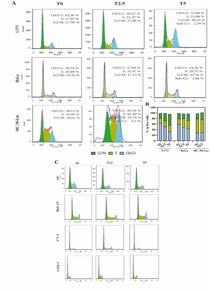
). The profiles were obtained by FACS Calibur flow cytometry (BD Science), and cell cycle progression was based on DNA quantification. Cells were modulated for 5 min, then collected at 48 h afterward, fixed and stained with the PI/RNase buffer (BD Pharmingen).
Fig. 5
Fig. 5
Evaluation of MEMP effects on cell shape and actin cytoskeleton organization. Cells were treated with MEMP for two minutes and immediately fixed for subsequent inspection by confocal microscopy with specific antibodies. (A) Fields of A9 and U373 cells stained with the indicated antibody and phalloidin and analyzed by confocal microscopy at 60x magnification. Scale bars, 20 μm. (B) Quantitative determination of the collapse of the actin cytoskeleton as determined by confocal microscopy (see Materials and Methods). Each dot represents a single cell (N, at least180 cells per condition). Statistics was performed using the Brown-Forsythe and Welch ANOVA and the Games-Howell´s multiple comparison tests. (C) Representative picture showing the colony forming ability of A9 and U373 cells at the MEMP regime used in A and B.
Fig. 6
Fig. 6
Evaluation of tumor forming capacity of modulated colon cancer cells in mice. Mouse MC-38-Luc adenocarcinoma cells were modulated by MEMP for 0, 1.5, and 2 min and then 0.5 × 106 control and modulated cells were injected per C57BL/6 mouse. (A) In vivo imaging quantification of tumor bioluminescence from day 0 (30 min post-inoculation) until day 19 (sacrifice of untreated cells-inoculated mice) of the first experiment. Inset: the same graph in log10 scale. Data were statistically analysed using a two-way ANOVA with Tukey post-hoc test. (B) IVIS images from day 0 until 19 days post-inoculation (radiance p/sec/cm2/sr color scale Min = 4.00e4, Max = 2.50e7) of the first in vivo experiment. (C) In vivo tumor progression from day 0 to day 5 post-inoculation based on the tumoral ratio to day 0 of all mice of the second experiment. Statistic was performed using a two-way ANOVA with Dunnett’s test. (D) IVIS images at day 0 and day 5 post-inoculation (radiance p/sec/cm2/sr color scale Min = 1.94e4, Max = 2.99e6) of the second experiment.

References

    1. Berg, H. et al. Bioelectromagnetic field effects on cancer cells and mice tumors. Electromagn. Biol. Med.29, 132–143. 10.3109/15368371003776725 (2010). - PubMed
    1. Williams, C. D., Markov, M. S., Hardman, W. E. & Cameron, I. L. Therapeutic electromagnetic field effects on angiogenesis and tumor growth. Anticancer Res.21, 3887–3891 (2001). - PubMed
    1. Yamaguchi, S., Ogiue-Ikeda, M., Sekino, M. & Ueno, S. Effects of pulsed magnetic stimulation on tumor development and immune functions in mice. Bioelectromagnetics27, 64–72. 10.1002/bem.20177 (2006). - PubMed
    1. Koh, E. K. et al. A 60-Hz sinusoidal magnetic field induces apoptosis of prostate cancer cells through reactive oxygen species. Int. J. Radiat. Biol.84, 945–955. 10.1080/09553000802460206 (2008). - PubMed
    1. Li, J., Ma, Y., Li, N., Cao, Y. & Zhu, Y. Natural static magnetic field-induced apoptosis in liver cancer cell. Electromagn. Biol. Med.33, 47–50. 10.3109/15368378.2013.783850 (2014). - PubMed

LinkOut - more resources