. 2024 Dec 3;14(1):30025.
doi: 10.1038/s41598-024-81102-w.

Dynamic changes in human brain connectivity following ultrasound neuromodulation

Affiliations

Dynamic changes in human brain connectivity following ultrasound neuromodulation

Cyril Atkinson-Clement et al. Sci Rep. .

Abstract

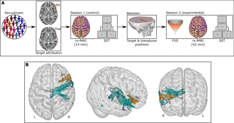
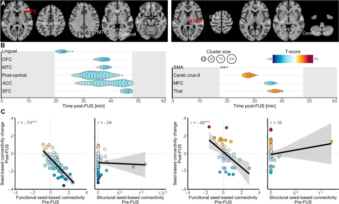
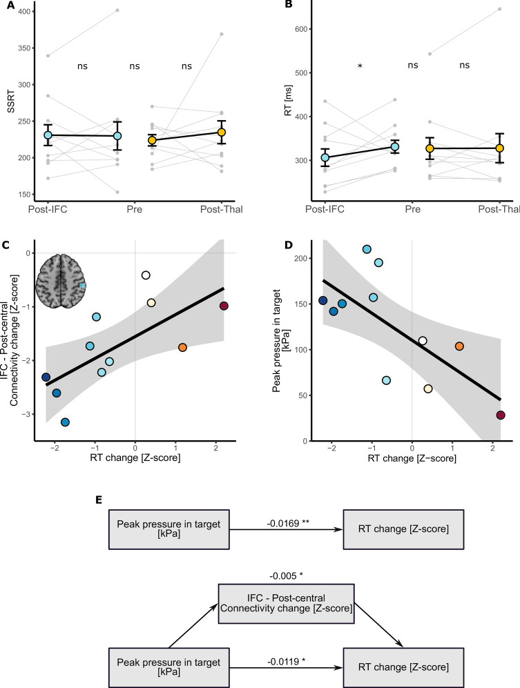
Non-invasive neuromodulation represents a major opportunity for brain interventions, and transcranial focused ultrasound (FUS) is one of the most promising approaches. However, some challenges prevent the community from fully understanding its outcomes. We aimed to address one of them and unravel the temporal dynamics of FUS effects in humans. Twenty-two healthy volunteers participated in the study. Eleven received FUS in the right inferior frontal cortex while the other 11 were stimulated in the right thalamus. Using a temporal dynamic approach, we compared resting-state fMRI seed-based functional connectivity obtained before and after FUS. We also assessed behavioural changes as measured with a task of reactive motor inhibition. Our findings reveal that the effects of FUS are predominantly time-constrained and spatially distributed in brain regions functionally connected with the directly stimulated area. In addition, mediation analysis highlighted that FUS applied in the right inferior cortex was associated with behavioural alterations which was directly explained by the applied acoustic pressure and the brain functional connectivity change we observed. Our study underscored that the biological effects of FUS are indicative of behavioural changes observed more than an hour following stimulation and are directly related to the applied acoustic pressure.

Keywords: Focused ultrasound stimulation; Motor inhibition; Non-invasive neuromodulation; Seed-based connectivity; Stop signal task; Whole brain.


Conflict of interest statement

Declarations. Competing interests: The authors declare no competing interests.

Figures

Fig. 1
Fig. 1
Study design. (A) 22 participants were recruited and then divided into two groups according to the brain target (IFC or Thal). The first session (“control”) consisted of an MRI protocol and a cognitive task. Next, the “between” section refers to the data preprocessing to identify the target and transducer positions. Finally, the second session (“experimental”) involved to the actual FUS followed by an MRI and a cognitive task. (B) Representation of the activated volume based on the acoustic simulations for all participants. Orange refers to the 11 participants with IFC-FUS whilst turquoise refers to the 11 participants with the Thalamus-FUS. IFC, inferior frontal cortex; L, Left; R, Right; rs-fMRI, resting-state functional MRI; SST, stop signal task; Thal, thalamus.
Fig. 2
Fig. 2
Functional connectivity changes following FUS. (A) Spatial locations of the significant clusters of seed-based connectivity changes following IFC-FUS (left) and Thalamus-FUS (right). The red sphere highlights the location of the target which was also used as seed. (B) Temporal locations of the significant clusters of seed-based connectivity changes following IFC-FUS (left) and Thalamus-FUS (right). The size of the dot reflects the size of the cluster (i.e., number of voxels) and the colours reflect the value of the peak at each moment (i.e., based on the T-score. A positive score indicates a significantly increased connectivity in comparison to the control condition, while a negative score indicates a decrease). (C) Correlations between the seed-based connectivity changes for each participant and cluster in the peak coordinates (in the 4 dimensions x, y, z, time) and the functional or structural seed-based connectivity for the IFC-FUS (left) and the Thalamus-FUS (right). ACC, anterior cingulate cortex; IFC, inferior frontal cortex; MFC, middle frontal cortex; MTC, middle temporal cortex; OFC, orbitofrontal cortex; SFC, superior frontal cortex; SMA, supplementary motor area; Thal, Thalamus.
Fig. 3
Fig. 3
Behavioural changes following FUS. (A) No reactive inhibition (as measured by the SSRT) was found neither between groups nor after FUS. (B) Decreased response time was observed after IFC-FUS while no change was found after Thalamus-FUS. (C) Decrease in response time was significantly correlated with the decreased functional connectivity between the IFC and the post-central cortex. (D) Decrease in response time was also significantly correlated with the peak pressure estimated in the IFC. (E) The direct effect of the peak pressure applied to the IFC on the decrease in response time (top) was significantly mediated by the decreased functional connectivity between the IFC and the post-central cortex (bottom). The values correspond to the β of each effect. IFC, inferior frontal cortex; Post, refers to the experimental session, following FUS; Pre, refers to the control session, without FUS; RT, response time; SSRT, stop signal reaction time; Thal. Thalamus; ns, not significant; *p < 0.05; **p < 0.01.

References

    1. Rabut, C. et al. Ultrasound technologies for imaging and modulating neural activity. Neuron108, 93–110. 10.1016/j.neuron.2020.09.003 (2020). - DOI - PMC - PubMed
    1. Legon, W. et al. A retrospective qualitative report of symptoms and safety from transcranial focused ultrasound for neuromodulation in humans. Sci. Rep.10, 5573. 10.1038/s41598-020-62265-8 (2020). - DOI - PMC - PubMed
    1. Blackmore, J., Shrivastava, S., Sallet, J., Butler, C. R. & Cleveland, R. O. Ultrasound neuromodulation: A review of results, mechanisms and safety. Ultrasound Med. Biol.45, 1509–1536. 10.1016/j.ultrasmedbio.2018.12.015 (2019). - DOI - PMC - PubMed
    1. Darmani, G. et al. Non-invasive transcranial ultrasound stimulation for neuromodulation. Clin. Neurophysiol.135, 51–73. 10.1016/j.clinph.2021.12.010 (2022). - DOI - PubMed
    1. Plaksin, M., Kimmel, E. & Shoham, S. Cell-Type-selective effects of intramembrane cavitation as a unifying theoretical framework for ultrasonic neuromodulation. Eneuro10.1523/ENEURO.0136-15.2016 (2016). - DOI - PMC - PubMed

LinkOut - more resources