. 2025 Jan;40(1):1-32.
doi: 10.1007/s12928-024-01044-y. Epub 2024 Dec 3.

Clinical expert consensus document on bailout algorithms for complications in percutaneous coronary intervention from the Japanese Association of Cardiovascular Intervention and Therapeutics

Affiliations

Clinical expert consensus document on bailout algorithms for complications in percutaneous coronary intervention from the Japanese Association of Cardiovascular Intervention and Therapeutics

Takayuki Ogawa et al. Cardiovasc Interv Ther. 2025 Jan.

Abstract

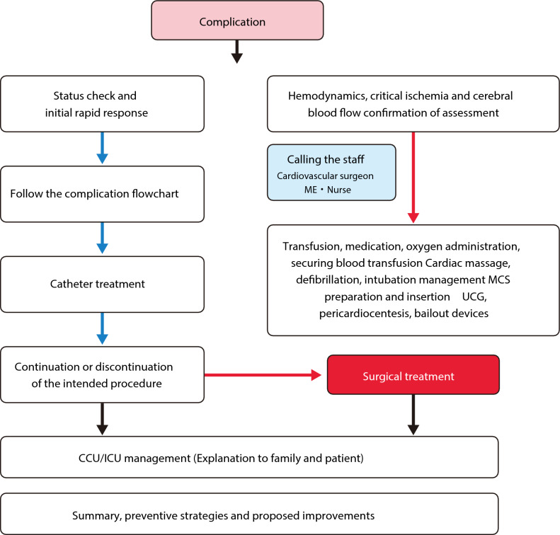
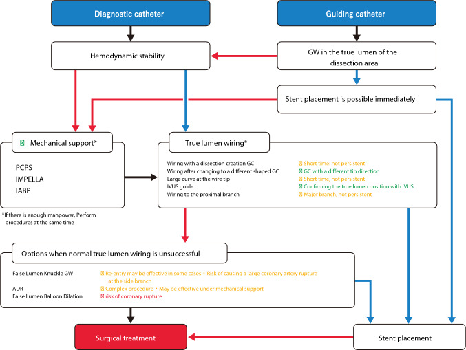
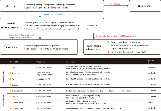
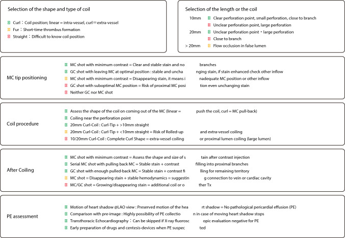
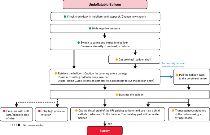
The efficacy and safety of percutaneous coronary intervention (PCI) for coronary artery disease has been established, and approximately 250,000 PCI procedures are performed annually in Japan. However, various complications including life-threatening complications can occur during PCI. Although several bailout procedures have been proposed to address complications during PCI, it is critically important for operators to manage each complication in real catheter rooms with confidence even in emergent situations. Standard bailout methods including specific techniques should be clarified as algorithms and shared with inexperienced operators as well as experienced operators. The Task Force of the Japanese Society for Cardiovascular Intervention and Therapeutics (CVIT) has developed the expert consensus document on bailout algorithms for complications in PCI.

Keywords: Bailout; Complication; Percutaneous coronary intervention.


Figures

Fig. 1
Fig. 1
Flowchart for PCI complications. (Green right arrow) Successful (YES). (Red right arrow) Unsuccessful (NO). (Black right arrow) Progress
Fig. 2
Fig. 2
Flowchart for severe coronary dissection caused by catheter. (Green right arrow) Successful (YES). (Red right arrow) Unsuccessful (NO). (Black right arrow) Progress. (Green circle) Recommended. (Yellow circle) Not recommended, but no other method is available, and successes have been reported. (Red circle) Not recommended. GW, guidewire; PCPS, percutaneous cardiopulmonary support; GC, guiding catheter; IABP, intra-aortic balloon pumping; IVUS, intravascular ultrasound; ADR, antegrade dissection re-entry
Fig. 3
Fig. 3
Flowchart for coronary perforation. MC, microcatheter
Fig. 4
Fig. 4
Coil placement techniques using curled coils
Fig. 5
Fig. 5
IVUS image of high-risk lesions for coronary rupture. IVUS image of a coronary rupture lesion (upper panels, calcified lesions; lower panels, fibrotic lesions). All lesions were highly eccentric and no plaques were observed on the opposite side
Fig. 6
Fig. 6
IVUS image of extravascular wiring. A IVUS image of extravascular wiring. B The white dotted area on the right is a space created outside the vessel, and the area indicated in red is hypoechoic compared with the healthy perivascular tissue, strongly suggesting an extravascular hematoma. The dotted yellow line indicates the original coronary artery
Fig. 7
Fig. 7
Flowchart for blowout-type coronary artery rupture
Fig. 8
Fig. 8
Procedure steps of double GC system/pericardiocentesis trouble
Fig. 9
Fig. 9
Flowchart for guidewire entrapment or fracture. CTO, chronic total occlusion; GEC, guide-extension catheter
Fig. 10
Fig. 10
The “Knuckle-twister” technique. This method retrieves a fractured or entrapped guidewire by forming the tip of the polymer-jacketed guidewire into a knuckle of 3–6 cm (A), advancing it slightly distal to the lost wire (B), and pulling it back while continuously drilling until the wire becomes completely entangled with the lost wire (D, E). This figure has been reprinted with permission (Leibundgut et al. [29])
Fig. 11
Fig. 11
Experimental image of the mechanisms of IVUS entrapment occurrence. Experimental image of IVUS entrapment after stent placement (A). Experimental image during the insertion of a small-diameter balloon or microcatheter (B). The device tip occluded the exit port, leaving no space for entrapment of the stent strut
Fig. 12
Fig. 12
Flowchart for imaging entrapment
Fig. 13
Fig. 13
How to cut and pull the drive shaft sheath. A An RA burr (1.25 mm) is inserted into a 6 Fr guide catheter via a Y-connector. B, C The drive shaft, drive shaft sheath, and RA wire are cut together near the advancer. D, E The drive shaft sheath was pulled back and removed. F After the drive shaft sheath is removed, the drive shaft remains in the same position. G, H A guidewire (0.014″) is passed through the guide catheter via an inserter and Y-connector. I A 2.5 × 15 mm conventional balloon easily passed through the guide catheter. This figure was reproduced with permission from Sakakura et al. [37]
Fig. 14
Fig. 14
Flowchart for burr entrapment. DS, drive shaft; DSS, drive shaft sheath; GW, guidewire
Fig. 15
Fig. 15
Flowchart for stent dislodgement. Stenting: if there is no obvious stent deformation, there is no significant disadvantage to implanting the stent at the site of stent loss, and if the guidewire is still inside the stent, stenting at the site of stent loss is an option. The surgeon can attempt to insert a small balloon (balloon size 1.5 mm × 10 mm) into the dislodged stent. If successful, the surgeon should increase the balloon size to match the vessel diameter at the implantation site. Stent retrieval. Small balloon method. The operator inserts a small-diameter balloon into the dislodged stent, dilates the balloon at the proximal site, and retrieves it. Snare method. A snare catheter can also be used to retrieve a dislodged stent through the guidewire inside the stent using Medtronic Goose Neck Snares. Small balloon/snare combination. If delivery of a snare is challenging, it can be mounted on a small-diameter balloon to reach the stent site. Multi-wire method. Two additional guidewires are added outside the stent and advanced into another branch on its peripheral side. Thereafter, the three guidewires are placed on a torquer and continuously turned in the same direction. When the stent starts to rotate, it should be moved proximally and retrieved while maintaining rotation. This method is not recommended because of the possibility of stent detachment from the multi-wire and low success rates
Fig. 16
Fig. 16
Small balloon with snare. Usually, a Goose Neck Snare is combined with a guidewire and a small-diameter balloon to facilitate reaching the dislodged stent
Fig. 17
Fig. 17
Homemade snare (Sumitsuji snare). Use GECs and balloons larger than 2 mm. A The normal-use wire is folded 20 mm from the tip of the monorail balloon as a reverse wire. The balloon diameter should be selected to ensure sufficient crimping of the guiding catheter during extension. B The tips of the folded wire and balloon are advanced close to the tip of the guiding extension catheter. CThe reverse portion of the wire is fixed. After balloon dilation, a snare loop is created by pushing the wire forward through the balloon
Fig. 18
Fig. 18
Flowchart for undeflatable balloon. The tip of the Heartrail II 5 Fr ST01 catheter (Terumo) is cut to expose the blade and inserted into the guiding catheter as a pediatric catheter. The catheter is placed close to the balloon, and its tip is pressed against the balloon to burst it. If the balloon is located distally, the catheter may be unable to reach it. Thus, the Y-connector should be replaced with a short hemostasis valve in advance. If the balloon cannot be ruptured by pressing the cut child’s catheter, the catheter is rotated slightly

References

    1. Yamaji K, Kohsaka S, Inohara T, Numasawa Y, Ando H, Wada H, Ishii H, Amano T, Miyata H, Ikari Y. Percutaneous coronary intervention during the COVID-19 pandemic in Japan: insights from the nationwide registration data. Lancet Reg Health West Pac. 2022;22: 100434. 10.1016/j.lanwpc.2022.100434. - PMC - PubMed
    1. Nakamura M, Yaku H, Ako J, Arai H, Asai T, Chikamori T, Daida H, Doi K, Fukui T, Ito T, et al. JCS/JSCVS 2018 guideline on revascularization of stable coronary artery disease. Circ J. 2022;86:477–588. 10.1253/circj.CJ-20-1282. - PubMed
    1. Giannini F, Candilio L, Mitomo S, Ruparelia N, Chieffo A, Baldetti L, Ponticelli F, Latib A, Colombo A. A practical approach to the management of complications during percutaneous coronary intervention. JACC Cardiovasc Interv. 2018;11:1797–810. 10.1016/j.jcin.2018.05.052. - PubMed
    1. Doll JA, Hira RS, Kearney KE, Kandzari DE, Riley RF, Marso SP, Grantham JA, Thompson CA, McCabe JM, Karmpaliotis D, et al. Management of percutaneous coronary intervention complications: algorithms from the 2018 and 2019 Seattle percutaneous coronary intervention complications conference. Circ Cardiovasc Interv. 2020;13: e008962. 10.1161/circinterventions.120.008962. - PubMed
    1. Klaudel J, Glaza M, Klaudel B, Trenkner W, Pawłowski K, Szołkiewicz M. Catheter-induced coronary artery and aortic dissections. A study of the mechanisms, risk factors, and propagation causes. Cardiol J. 2022. 10.5603/CJ.a2022.0050. - PMC - PubMed

Publication types

MeSH terms

LinkOut - more resources