Review
. 2024 Dec 3;22(1):581.
doi: 10.1186/s12964-024-01942-x.

Complexin regulation of synaptic vesicle release: mechanisms in the central nervous system and specialized retinal ribbon synapses

Affiliations
Review

Complexin regulation of synaptic vesicle release: mechanisms in the central nervous system and specialized retinal ribbon synapses

Yun-Zhi Li et al. Cell Commun Signal. .

Abstract

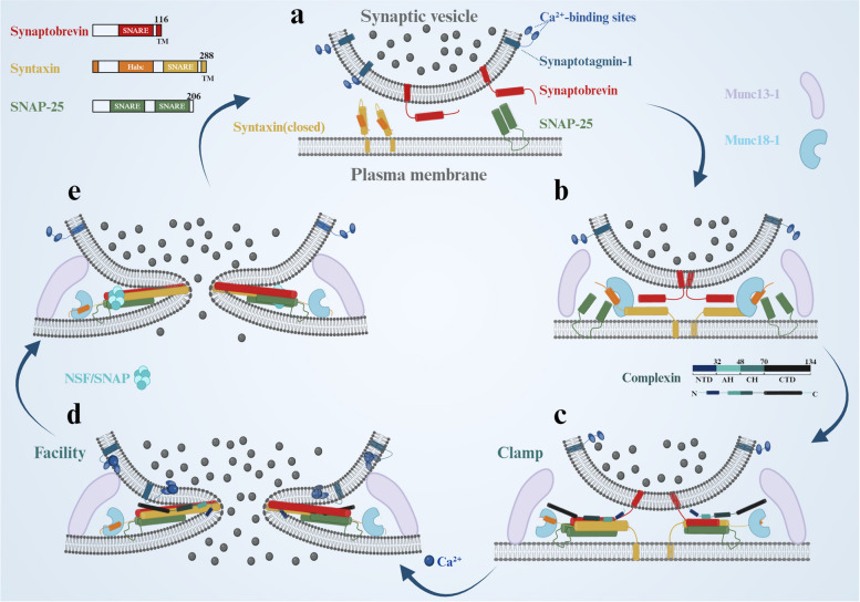
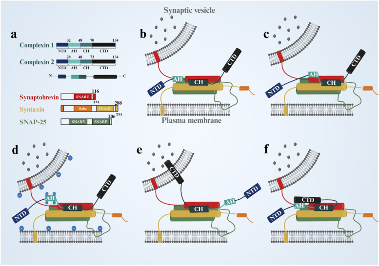
Synaptic ribbons, recognized for their pivotal role in conveying sensory signals in the visual pathway, are intricate assemblages of presynaptic proteins. Complexin (CPX) regulates synaptic vesicle fusion and neurotransmitter release by modulating the assembly of the soluble NSF attachment protein receptor (SNARE) complex, ensuring precise signal transmission in the retina and the broader central nervous system (CNS). While CPX1 or CPX2 isoforms (CPX1/2) play crucial roles in classical CNS synapses, CPX3 or CPX4 isoforms (CPX3/4) specifically regulate retinal ribbon synapses. These isoforms are essential for sustaining synaptic plasticity related to light signaling, adapting to changes in circadian rhythms, and dynamically regulating visual function under varying light conditions. This review explores the regulation of synaptic vesicle release by CPX in both the CNS and retinal ribbon synapses, with a focus on the mechanisms governing CPX3/4 function in the retina. Additionally, by reviewing the role of CPX and ribbon synapse dysfunction in non-retinal diseases, we further hypothesize the potential mechanisms of CPX in retinal diseases and propose therapeutic strategies targeting CPX to address retinal and CNS disorders associated with synaptic dysfunction.

Keywords: Central nervous system; Complexin; Photoreceptor synapse; Retina; Ribbon synapse; SNARE complex.


Conflict of interest statement

Declarations. Ethics approval and consent to participate: Not applicable. Consent for publication: Not applicable. Competing interests: The authors declare no competing interests.

Figures

Fig. 1
Fig. 1
SNARE complex-mediated synaptic vesicle fusion cycle. a The distribution of synaptobrevin, synaptotagmin-1, syntaxin, and SNAP-25 on the presynaptic and plasma membranes is depicted. The Habc domain of syntaxin (orange) binds to the SNARE motif (H3 domain) (yellow), thereby maintaining the closed state of syntaxin. b Munc18-1 and Munc13-1 collaboratively regulate the transition of Syntaxin-1 to its open state, facilitating the initiation of SNARE complex assembly. c The partial assembly of the SNARE complex promotes the close contact between the vesicle and the plasma membrane. Meanwhile, Complexin inserts into the groove between synaptobrevin and syntaxin, exhibiting its clamp function to inhibit premature fusion. d The interaction between Ca2+ and Synaptotagmin-1 triggers the binding of NTD of CPX to the fully assembled SNARE complex, facilitating its association with the plasma membrane. This interaction drives the fusion of phospholipid molecules within the membrane, ultimately leading to the formation of a fusion pore, highlighting Complexin's facilitative role. e NSF/SNAP disassembles the SNARE complex, thereby allowing the vesicles to be recycled and reused in the process of release
Fig. 2
Fig. 2
CPX inhibits the assembly of the SNARE complex. a Schematic representation of different domains within CPX and the SNARE complex proteins. Numbers indicate the amino acid positions at the termination points of the respective domains. (b, c, d) Models illustrating the mechanism of AH in directly inhibiting SNARE complex assembly: b The AH inhibits SNARE complex assembly by inserting itself into the SNARE complex. c The AH binds to synaptobrevin, blocking the assembly of the SNARE complex. d The AH increases electrostatic repulsion between membranes, thereby inhibiting SNARE complex assembly. e, f Models illustrating the indirect inhibition of SNARE complex assembly by the CTD: e The CTD anchors to highly curved vesicle membranes, guiding the CH to insert into the groove between synaptobrevin and syntaxin, thereby indirectly preventing the complete assembly of the SNARE complex at its C-terminal. f The CTD folds back onto the AH, enhancing the functional activity of the AH and indirectly blocking the complete assembly of the SNARE complex. NT = N-terminal domain, AH = accessory helix, CH = central helix, and CTD = C-terminal domain

References

    1. Kaeser PS, Regehr WG. Molecular mechanisms for synchronous, asynchronous, and spontaneous neurotransmitter release. Annu Rev Physiol. 2014;76:333–63. - PMC - PubMed
    1. Hua Z, Leal-Ortiz S, Foss SM, Waites CL, Garner CC, Voglmaier SM, et al. v-SNARE composition distinguishes synaptic vesicle pools. Neuron. 2011;71(3):474–87. - PMC - PubMed
    1. Reim K, Wegmeyer H, Brandstätter JH, Xue M, Rosenmund C, Dresbach T, et al. Structurally and functionally unique complexins at retinal ribbon synapses. J Cell Biol. 2005;169(4):669–80. - PMC - PubMed
    1. Thoreson WB, Dacey DM. Diverse Cell Types, Circuits, and Mechanisms for Color Vision in the Vertebrate Retina. Physiol Rev. 2019;99(3):1527–73. - PMC - PubMed
    1. Townes-Anderson E, Halasz E, Wang W, Zarbin M. Coming of Age for the Photoreceptor Synapse. Invest Ophthalmol Vis Sci. 2021;62(12):24. - PMC - PubMed

MeSH terms

Substances

LinkOut - more resources