. 2025 Feb;100(2):342-345.
doi: 10.1002/ajh.27545. Epub 2024 Dec 3.

Feasibility of a Biomarker-Based Screening for Pre-Symptomatic AL Amyloidosis in Patients With Intermediate/High-Risk MGUS

Affiliations

Feasibility of a Biomarker-Based Screening for Pre-Symptomatic AL Amyloidosis in Patients With Intermediate/High-Risk MGUS

Silvia Mangiacavalli et al. Am J Hematol. 2025 Feb.

Abstract

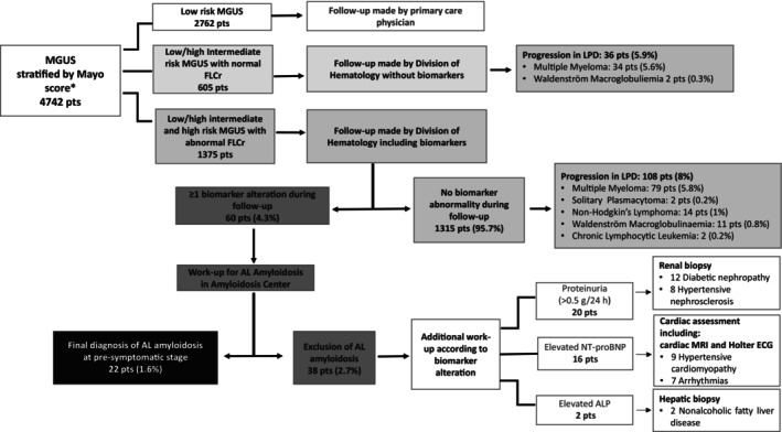
Biomarker-based screening enables early detection of AL amyloidosis in intermediate/high-risk MGUS patients. Our study shows that identifying pre-symptomatic AL through biomarker longitudinal monitoring allows early treatment, leading to significant organ function recovery.

Keywords: AL amyloidosis; MGUS; biomarkers; screening.


Conflict of interest statement

SM has received honoraria from Bristol‐Myers Squibb, Sanofi, AMGEN, GSK, Takeda, Pfizer, and Janssen, has served on the advisory boards for Sanofi, Takeda, Bristol‐Myers Squibb, Pfizer, and Janssen; GP received honoraria from Pfizer, Serbia, and Siemens and served on advisory boards Alexion, Argobio, GSK, Janssen, and Prothena. PM has received honoraria from Janssen, Pfizer (also research support), Prothena, and Siemens and served on the advisory board for Siemens. LA received honoraria from EUSA Pharma, and Novartis, and served on advisory boards and consultations from Roche, incite, EUSA Pharma, Kite/Gilead, Novartis, and Morphosys.

Figures

FIGURE 1
FIGURE 1
Flow chart of patient disposition during the study period. Baseline evaluation at the time of first detection of monoclonal component (MC) included complete blood count, serum calcium, serum creatinine, serum protein electrophoresis, serum and urine immunofixation, free light chain quantification and ratio (FLCr) and 24‐h proteinuria. Diagnosis of MGUS was established according to International Myeloma Working Group (IMWG) criteria. In cases of low‐risk Monoclonal Gammopathy of Undetermined Significance (MGUS) according to Mayo risk stratification (i.e., MC ≤ 1,5 g/dL, Immunoglobulin G [IgG] type, normal FLCr), no further tests were performed and patients were monitored after 6 months at the Division of Hematology and then, if stable, they were referred back to primary care physician for follow‐up. In patients with non‐low‐risk MGUS according to the Mayo risk score, baseline evaluation included skeletal imaging (low‐dose whole‐body computed tomography) along with bone marrow evaluation, to rule out multiple myeloma. Follow‐up of these patients was carried out at the Division of Hematology, firstly after 6 months and then every year in case of no significant change. Follow‐up tests included: complete blood count, serum calcium, serum creatinine, serum protein electrophoresis, 24‐h proteinuria, and FLC analysis. In intermediate (low/high) as well as high‐risk MGUS with altered FLCr, follow‐up tests were integrated by assessment of biomarkers of amyloid organ involvement (N‐terminal pro‐type‐B natriuretic peptide [NT‐proBNP], 24‐h proteinuria, and alkaline phosphatase). All MGUS patients with at least one abnormal biomarkers according to predefined cut‐off values (suspect kidney involvement: 24‐h proteinuria > 0.5 g/24 h, predominantly albumin; suspect liver involvement: Alkaline phosphatase concentration > 1.5 times the institutional upper limit of normal; suspect cardiac involvement: NT‐proBNP concentration > 332 ng/L) underwent a new follow‐up within three months, in case of confirmed biomarker alteration a multistep diagnostic workup, in particular abdominal fat biopsy, was performed at the Amyloidosis Research and Treatment Center. The diagnosis of AL amyloidosis was biopsy‐based and included amyloid typing by immuno‐electron microscopy or mass spectrometry. Patients with negative abdominal fat biopsy underwent minor salivary gland or organ biopsy as appropriate. Amyloid organ involvement was defined and graded according to the International Society of Amyloidosis guidelines. In cases in whom amyloidosis was excluded, diagnostic algorithm included additional multidisciplinary specific workup performed in accordance with type of biomarker alteration identified, to rule out alternative non‐hematological morbidities. MGUS, Monoclonal gammopathy of undetermined significance; FLCr, Free Light Chain ratio; Ps, patients; LPD, Lymphoproliferative Disorders; AL, Light Chain Amyloidosis; NT‐proBNP, N‐terminal pro‐type‐B Natriuretic Peptide; ALP, Alkaline phosphatase; MRI, Magnetic Resonance Imaging; ECG, electrocardiogram; h, hour.* According to the Mayo risk model, low‐risk MGUS meets all the following characteristics: Monoclonal Component < 1.5 g/dL, Immunoglobulin G subtype, normal FLCr (0.26–1.65), low‐intermediate risk MGUS had any one factor abnormal, high‐intermediate risk MGUS had any 2 factors abnormal, high‐risk MGUS had all three factors abnormal.

References

    1. Kyle R. A., Larson D. R., Therneau T. M., et al., “Long‐Term Follow‐Up of Monoclonal Gammopathy of Undetermined Significance,” New England Journal of Medicine 378, no. 3 (2018): 241–249, 10.1056/NEJMoa1709974. - DOI - PMC - PubMed
    1. Palladini G. and Merlini G., “How I Treat AL Amyloidosis,” Blood 139, no. 19 (2022): 2918–2930, 10.1182/blood.2020008737. - DOI - PubMed
    1. Weiss B. M., Hebreo J., Cordaro D. V., et al., “Increased Serum Free Light Chains Precede the Presentation of Immunoglobulin Light Chain Amyloidosis,” Journal of Clinical Oncology 32, no. 25 (2014): 2699–2704, 10.1200/JCO.2013.50.0892. - DOI - PMC - PubMed
    1. Sharpley F. A., Fontana M., Martinez‐Naharro A., et al., “Cardiac Biomarkers Are Prognostic in Systemic Light Chain Amyloidosis With no Cardiac Involvement by Standard Criteria,” Haematologica 105 (2019): 1405–1413, 10.3324/haematol.2019.217695. - DOI - PMC - PubMed
    1. Gonzalez‐Lopez E., Escobar‐Lopez L., Obici L., et al., “Prognosis of Transthyretin Cardiac Amyloidosis Without Heart Failure Symptoms,” JACC CardioOncol 4, no. 4 (2022): 442–454, 10.1016/j.jaccao.2022.07.007. - DOI - PMC - PubMed

MeSH terms

LinkOut - more resources