. 2024 Nov 19:15:1481181.
doi: 10.3389/fimmu.2024.1481181. eCollection 2024.

Alpha-1-antitrypsin as novel substrate for S. aureus' Spl proteases - implications for virulence

Affiliations

Alpha-1-antitrypsin as novel substrate for S. aureus' Spl proteases - implications for virulence

Franziska Scherr et al. Front Immunol. .

Abstract

Background: The serine protease like (Spl) proteases of Staphylococcus aureus are a family of six proteases whose function and impact on virulence are poorly understood. Here we propose alpha-1-antitrypsin (AAT), an important immunomodulatory serine protease inhibitor as target of SplD, E and F. AAT is an acute phase protein, interacting with many proteases and crucial for prevention of excess tissue damage by neutrophil elastase during the innate immune response to infections.

Methods: We used MALDI-TOF-MS to identify the cleavage site of Spl proteases within AAT's reactive center loop (RCL) and LC-MS/MS to quantify the resulting peptide cleavage product in in vitro digestions of AAT and heterologous expressed proteases or culture supernatants from different S. aureus strains. We further confirmed proteolytic cleavage and formation of a covalent complex with Western Blots, investigated AAT's inhibitory potential against Spls and examined the NETosis inhibitory activity of AAT-Spl-digestions.

Results: SplD, E and F, but not A or B, cleave AAT in its RCL, resulting in the release of a peptide consisting of AAT's C-terminal 36 amino acids (C36). Synthetic C36, as well as AAT-SplD/E/F-digestions exhibit NETosis inhibition. Only SplE, but not D or F, was partly inhibited by AAT, forming a covalent complex.

Conclusion: We unraveled a new virulence trait of S. aureus, where SplD/E/F cleave and inactivate AAT while the cleavage product C36 inhibits NETosis.

Keywords: AAT; C-terminal Alpha-1-Antitrypsin peptides; CAAPs; NETosis; Staphylococcus aureus; host-pathogen interaction; virulence.


Conflict of interest statement

MK is an inventor of a patent application covering the utilized LC-MS/MS method as a tool for characterizing systemic inflammation applicant: Jena University Hospital; inventors: Arite Bigalke and MK; published as EP4224163A1. Jena University Hospital is owner of a patent related to methods determining the origin of an infection EP3239712: granted; inventors: MK, Diana Schmerler. A patent covering the initial identification of C42 was granted as well published as CN104204808B, JP6308946B2, US10712350B2, EP2592421B1, EP2780719B1; owner: Jena University Hospital; Inventors: MK, Diana Schmerler, Thomas Deufel, Frank Brunkhorst. The remaining author declares that the research was conducted in the absence of any commercial or financial relationships that could be construed as a potential conflict of interest.

Figures

Figure 1
Figure 1
Overview of Alpha-1-Antitrypsin’s C-terminal cleavage sites and reaction mechanism (A) Depicts the C-terminal amino acid sequence of Alpha-1-Antitrypsin (AAT) starting at position 368, with the reactive center loop marked in blue. Cleavage sites of both exogenous as well as endogenous proteases are indicated above or below the sequence, respectively (, –, –49). Known cleavage sites of proteases from S. aureus are highlighted in dark red. (B) shows the reaction pathway of AAT. Upon interaction with any serine protease E, a non-covalent Michaelis complex [AAT-E] is formed. Cleavage of AAT by the protease leads to the formation of an intermediate, covalent acyl-enzyme complex [AAT-E]’, which can either be stabilized into a covalent complex with inactivated protease [AAT-E]* by AAT’s conformational change. Alternatively, the intermediate complex can also dissociate into a cleaved AAT, AAT* and the regenerated protease E. Over time, the covalent complex [AAT-E]* can slowly dissociate into AAT* and E. AAT, alpha-1-antitrypsin; k, rate constant.
Figure 2
Figure 2
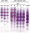
Alpha-1-Antitrypsin cleavage by Spl proteases from S. aureus Pure AAT or mixtures of AAT and indicated proteases were incubated for 3 h at 37°C before analysis by isoelectric focusing. Lanes 1-3 depict different pure AAT isoforms (normal PI*MM, pathologic PI*MS, pathologic PI*MZ genotype [4]). Upon incubation of plasma-derived AAT with SplA or B (lanes 4-5), a similar pattern can be observed. In contrast, SplD, E and F (lanes 6-8) produce a markedly different pattern, indicating cleavage of AAT. Lane 9 displays the impact of MMP7, which is known to cleave AAT at several sites, and shows a distinct pattern compared to the Spl proteases. AAT, alpha-1-antitrypsin; MMP, matrix metalloprotease.
Figure 3
Figure 3
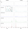
MALDI-TOF-MS spectra of AAT and AAT-Spl digestions Spectra display the peptide mass range (1700-4500 m/z) on the left and protein mass range on the right (17000-55000 m/z) of the average of n=3 independent experiments. The inlet shows a zoomed-in portion of the respective spectra to better distinguish the signals. AAT or AAT plus respective Spl protease was incubated in PBS at 37°C for 21 h before sample preparation and measurements as described in Materials and Methods. Single charged native or digested AAT can be seen around 52000 or 47000 m/z, respectively. Spl proteases are visible at approximately 22500 m/z. While SplD_mut did not show any digestion of AAT, SplD, E and F all cleaved AAT at the same site. AAT, alpha-1-antitrypsin.
Figure 4
Figure 4
Proteolytic cleavage of AAT by Spl proteases or S. aureus culture supernatants 1.5 mg/mL AAT was incubated with either purified Spl proteases D, D_mut, E and F at 1:10 molar ratio (A) or with supernatants from different S. aureus strains (B) harboring different spl operon compositions (C). In (A), values represent the mean ± standard deviation of three independent replicates. Concentration levels in (B) are normalized to the total protein amount of the supernatants and represent the mean of two independent experiments. Strains in (B) are grouped by either containing any combination of splD, splE and splF or none thereof. AAT, alpha-1-antitrypsin.
Figure 5
Figure 5
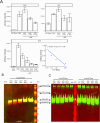
Effect of AAT on Spl protease activity (A) Native Spls or Spls pretreated with indicated molar ratios of AAT were incubated with AMC-conjugated substrates as described in the methods section. Bar graphs show mean fluorescence intensities ± standard deviation after 30 min (for SplD and SplE) or 60 min (SplF) reaction time of two or three experiments performed in duplicates. For SplE, the residual activity was plotted against the inhibitor-enzyme ratio (I-E-Ratio) to determine the partition ratio (x-axis intercept). (B, C) display Western Blots of AAT or AAT-Spl digestions with indicated protein amounts per lane. In (B), a mix of two sets of primary and secondary antibodies was used to detect the C-terminus of AAT as green and the N-terminus as red fluorescence. As the C-terminus is cleaved off by SplD-F, the cleaved AAT only contains the native N-terminus and is therefore only detected in the red channel. In (C), only one set of antibodies was used to detect the N-terminus of AAT. Native and cleaved AAT therefore are both detected in the green channel. AAT, alpha-1-antitrypsin; AMC, 7-Amino-4-methylcoumarin.
Figure 6
Figure 6
NETosis inhibition by AAT and AAT-Spl digestions Isolated neutrophils from healthy donors were preincubated with respective proteins or peptides with subsequent NETosis induction by PMA. Cells were monitored for 4 h and the percentage of NETotic cells was determined by automated image processing. The percentage of NETotic cells over time is displayed as mean +/- standard deviation of three biological replicates with two technical replicates each. Comparing synthetic C36 to AAT or AAT-Spl digestions to the respective pure proteases, a moderate to strong NETosis inhibition can be observed. AAT, alpha-1-antitrypsin; PMA, Phorbol 12-myristate 13-acetate.

References

    1. Hu L, Umeda A, Kondo S, Amako K. Typing of Staphylococcus aureus colonising human nasal carriers by pulsed-field gel electrophoresis. J Med Microbiol. (1995) 42:127–32. doi: 10.1099/00222615-42-2-127 - DOI - PubMed
    1. Nouwen JL, Ott A, Kluytmans-Vandenbergh MFQ, Boelens HAM, Hofman A, van Belkum A, et al. . Predicting the staphylococcus aureus nasal carrier state: derivation and validation of a “Culture rule. Clin Infect Dis. (2004) 39:806–11. doi: 10.1086/423376 - DOI - PubMed
    1. Tong SY, Davis JS, Eichenberger E, Holland TL, Fowler VG, Jr. Staphylococcus aureus infections: epidemiology, pathophysiology, clinical manifestations, and management. Clin Microbiol Rev. (2015) 28:603–61. doi: 10.1128/CMR.00134-14 - DOI - PMC - PubMed
    1. Weiner LM, Webb AK, Limbago B, Dudeck MA, Patel J, Kallen AJ, et al. . Antimicrobial-resistant pathogens associated with healthcare-associated infections: summary of data reported to the national healthcare safety network at the centers for disease control and prevention, 2011-2014. Infect Control Hosp Epidemiol. (2016) 37:1288–301. doi: 10.1017/ice.2016.174 - DOI - PMC - PubMed
    1. Thammavongsa V, Kim HK, Missiakas D, Schneewind O. Staphylococcal manipulation of host immune responses. Nat Rev Microbiol. (2015) 13:529–43. doi: 10.1038/nrmicro3521 - DOI - PMC - PubMed

MeSH terms

LinkOut - more resources