. 2024 Nov 19:15:1454102.
doi: 10.3389/fimmu.2024.1454102. eCollection 2024.

Stimulation of microneedles alleviates pathology of Parkinson's disease in mice by regulating the CD4+/CD8+ cells from the periphery to the brain

Affiliations

Stimulation of microneedles alleviates pathology of Parkinson's disease in mice by regulating the CD4+/CD8+ cells from the periphery to the brain

Jin Hee Kim et al. Front Immunol. .

Abstract

Introduction: Immune dysfunction is a major cause of neuroinflammation and accelerates the progression of Parkinson's disease (PD). Numerous studies have shown that stimulation of specific acupuncture points (acupoints) can ameliorate PD symptoms. The purpose of this study was to investigate whether attaching microneedles to acupoints would improve PD pathology by recovering immune dysfunction.

Methods: The PD mouse model was induced by intrastriatal injection of 6-hydroxydopamine (6-OHDA), and microneedle patches (MPs) or sham patches (SPs) were attached to GB20 and GB34, representative acupoints for treating PD for 14 days.

Results: First, the behavioral experiment showed that motor disorders induced by 6-OHDA were significantly improved by MP. Simultaneously, 6-OHDA-induced dopaminergic neuronal death and brain neuroinflammation decreased. Conversely, SP had no effect on behavioral disorders, neuronal death, or neuroinflammation. Measurement results from flow cytometry of immune cells in the brain and blood revealed a disruption in the CD4+/CD8+ ratio in the 6-OHDA group, which was significantly restored in the MP group. The brain mRNA expression of cytokines was significantly increased in the 6-OHDA group, which was significantly decreased by MP.

Discussion: Overall, our results suggest that the attachment of MPs to GB20 and GB34 is a new method to effectively improve the pathology of PD by restoring peripheral and brain immune function.

Keywords: Parkinson’s disease; acupuncture point; microneedle; neuroinflammation; peripheral immune.


Conflict of interest statement

Authors NYY, SL, and DHJ were employed by the company Raphas Co. Ltd. The remaining authors declare that the research was conducted in the absence of any commercial or financial relationships that could be construed as a potential conflict of interest.

Figures

Figure 1
Figure 1
Overview of experimental procedure. Attachment points of patches on a mouse (A) and overall experimental schedule (B).
Figure 2
Figure 2
Effects of MPs attached to acupoints on PD symptoms in 6-OHDA-injected mice. Behavioral disorders were assessed using the OFT, pole test, rotarod test, and cylinder test. Representative images of the OFT are shown in (A), track length (B), center zone duration time (C), number of center zone entries (D), and velocity (E) were measured. T-turn and T-LA in the pole test (F, G), latency time in the rotarod test (H), and impaired paw usage rate in the cylinder (I) test were measured. Values are given as the mean ± S.E.M. Data were analyzed by One-way ANOVA followed by post hoc Dunnett’s multiple comparisons test. *p < 0.05, **p < 0.01 and ***p < 0.001 compared to the 6-OHDA group.
Figure 3
Figure 3
Effects of MPs attached to acupoints on dopaminergic neurons in ST and SN of 6-OHDA-injected mice. Representative images of DAT in ST (scale bar = 500 μm) and TH in ST (scale bar = 500 μm) and SN (scale bar = 200 μm) are shown in (A). Graphs were represented as optical density of DAT-immunoreactivity in ST (B), TH-immunoreactivity in ST (C) and SN (D). Representative band images of TH, Bcl-2, Bax, Nrf2, and HO-1 in SN are shown in (E). Graphs were represented as expression of TH (F), Bcl-2/Bax ratio (G), Nrf2 (H), and HO-1 (I) in SN and dopamine content in ST (J). Values are given as the mean ± S.E.M. Data were analyzed by One-way ANOVA followed by post hoc Dunnett’s multiple comparisons test or Student’s t-test. *p < 0.05, **p < 0.01 and ***p < 0.001 compared to the 6-OHDA group; $p < 0.05 using student’s t-test.
Figure 4
Figure 4
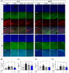
Effects of MPs attached to acupoints on expression of Iba-1 and GFAP in ST and SN of 6-OHDA-injected mice. Representative images of Iba-1 and GFAP in SN and ST (scale bar = 200 μm) are shown in (A). Graphs were represented as % Iba-1+ area in SN (B) and ST (D). Graphs were represented as % GFAP+ area in SN (C) and ST (E). Values are given as the mean ± S.E.M. Data were analyzed by One-way ANOVA followed by post hoc Dunnett’s multiple comparisons test or Student’s t-test. *p < 0.05, **p < 0.01 and ***p < 0.001 compared to the 6-OHDA group using One-way ANOVA; $p < 0.05 using student’s t-test.
Figure 5
Figure 5
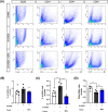
Effects of MPs attached to acupoints on % of B220, CD3+, CD4+, and CD8+ cells in the brain of 6-OHDA-injected mice. Representative flow cytometry plots of B220, CD3+, CD4+ and CD8+ cells in brain are shown in (A). Graphs were represented as % of B220 (B), CD3+ (C) and CD4+/CD8+ ratio (D) in brain. Values are given as the mean ± S.E.M. Data were analyzed by One-way ANOVA followed by post hoc Dunnett’s multiple comparisons test or Student’s t-test. **p < 0.01 and ***p < 0.001 compared to the 6-OHDA group using One-way ANOVA; $p < 0.05 using student’s t-test.
Figure 6
Figure 6
Effects of MPs attached to acupoints on mRNA expressions of cytokines in the brain of 6-OHDA-injected mice. The mRNA expressions of IL-6, IL-1β, IL-2, IL-4 and IL-17a in ST (A–E) and SN (F–J) were measured by qRT-PCR. Values are given as the mean ± S.E.M. Data were analyzed by One-way ANOVA followed by post hoc Dunnett’s multiple comparisons test. *p < 0.05 and **p < 0.01 compared to the 6-OHDA group using One-way ANOVA.
Figure 7
Figure 7
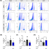
Effects of MPs attached to acupoints on % of B220, CD3+, CD4+ and CD8+ cells in the blood of 6-OHDA-injected mice. Representative flow cytometry plots of B220, CD3+, CD4+ and CD8+ cells in blood are shown in (A). Graphs were represented as % of B220 (B), CD3+ (C) and CD4+/CD8+ ratio (D) in blood. Values are given as the mean ± S.E.M. Data were analyzed by One-way ANOVA followed by post hoc Dunnett’s multiple comparisons test or Student’s t-test. *p < 0.05 compared to the 6-OHDA group using One-way ANOVA; $$p < 0.01 using student’s t-test.

References

    1. Barros-Santos T, Clarke G. Gut-initiated neuroprotection in Parkinson's disease: When microbes turn the tables in the battle against neuroinflammation. Brain Behav Immun. (2023) 108:350–2. doi: 10.1016/j.bbi.2022.12.016 - DOI - PubMed
    1. Huh E, Choi JG, Choi Y, Ju IG, Noh D, Shin DY, et al. . 6-shogaol, an active ingredient of ginger, improves intestinal and brain abnormalities in proteus mirabilis-induced parkinson's disease mouse model. Biomol Ther (Seoul). (2023) 31:417–24. doi: 10.4062/biomolther.2023.098 - DOI - PMC - PubMed
    1. Morris HR, Spillantini MG, Sue CM, Williams-Gray CH. The pathogenesis of Parkinson's disease. Lancet. (2024) 403:293–304. doi: 10.1016/S0140-6736(23)01478-2 - DOI - PubMed
    1. Balestrino R, Schapira AHV. Parkinson disease. Eur J Neurol. (2020) 27:27–42. doi: 10.1111/ene.14108 - DOI - PubMed
    1. Tansey MG, Wallings RL, Houser MC, Herrick MK, Keating CE, Joers V. Inflammation and immune dysfunction in Parkinson disease. Nat Rev Immunol. (2022) 22:657–73. doi: 10.1038/s41577-022-00684-6 - DOI - PMC - PubMed