Review
. 2024 Oct 28;27(12):111268.
doi: 10.1016/j.isci.2024.111268. eCollection 2024 Dec 20.

Autophagy in reproduction and pregnancy-associated diseases

Affiliations
Review

Autophagy in reproduction and pregnancy-associated diseases

Asmita Singh et al. iScience. .

Abstract

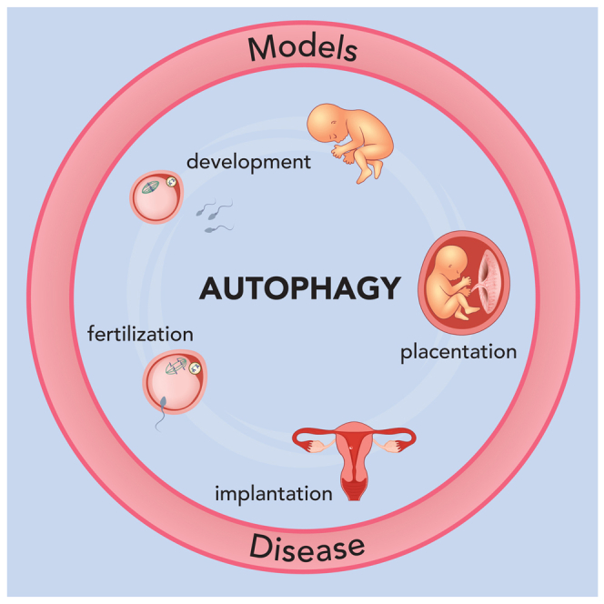
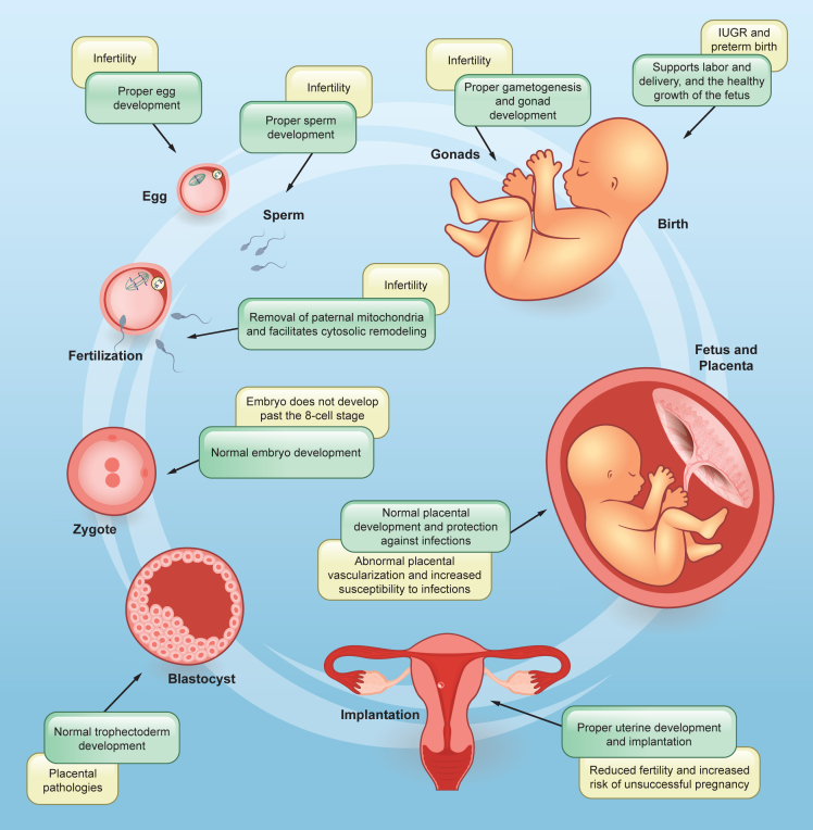
As advantageous as sexual reproduction is during progeny generation, it is also an expensive and treacherous reproductive strategy. The viviparous eukaryote has evolved to survive stress before, during, and after pregnancy. An important and conserved intracellular pathway for the control of metabolic stress is autophagy. The autophagy process occurs in multiple stages through the coordinated action of autophagy-related genes. This review summarizes the evidence that autophagy is an integral component of reproduction. Additionally, we discuss emerging in vitro techniques that will enable cellular and molecular studies of autophagy and its associated pathways in reproduction. Finally, we discuss the role of autophagy in the pathogenesis and progression of several pregnancy-related disorders such as preterm birth, preeclampsia, and intra-uterine growth restriction, and its potential as a therapeutic target.

Keywords: cell biology; molecular biology; pathophysiology; physiology.


Conflict of interest statement

All authors declare no competing interests.

Figures

None
Graphical abstract
Figure 1
Figure 1
Molecular players in the autophagy pathway Schematic overview of the mechanism of autophagy. (A) Cellular stress results in the formation of pre-autophagosomal structures. Autophagy initiation involves the mTOR inhibition and activation of the ULK complex, which recruits class III phosphatidylinositol 3-kinase complex C1 (PI3KC3-C1) to the phagophore formation site. This lipid kinase complex is responsible for phosphatidylinositol 3-phosphate (PI3P) generation on ATG9 vesicles, the now speculated membranous seed for phagophore formation. WIPI proteins bind to PI3P and subsequently recruit ATG2, which controls phospholipid trafficking into the growing phagophore from other membrane sources, mainly the endoplasmic reticulum. (B) The growing phagophore membrane is decorated with the ATG8/LC3 family of proteins and cargo receptors by a ubiquitin-conjugation-like machinery. LC3 is first processed from its precursor form to LC3-I through cleavage by ATG4, exposing a C-terminal glycine. LC3-I is then lipidated with phosphatidylethanolamine to form LC3-II, which integrates into autophagosomal membranes. (C) Sequestration of selectively ubiquitin-tagged or non-selective cargo recruitment into the expanding phagophore. This is followed by the sealing of the phagophore membrane by ESCRT machinery. (D) A fully formed double membrane autophagosome fuses with a lysosome to assist in the degradation, recycling, and secretion of all the encapsulated material.
Figure 2
Figure 2
Schematic of gametogenesis The formation of an embryo after fertilization requires the proper development of a healthy egg and sperm in the process of gametogenesis. (A) The generation of sperm in males is known as spermatogenesis. The diploid primary spermatocytes arise from diploid spermatogonia which undergo two meiotic divisions to form haploid spermatozoa. First meiotic division produces two haploid secondary spermatocytes, each of which undergo second meiotic division to form two haploid spermatids. These spermatids acquire flagella to mature into spermatozoa. (B) In females, the formation of oocytes is known as oogenesis. The diploid primary oocyte undergoes two rounds of meiotic divisions to form a haploid secondary oocyte and a polar body. The primary oocyte is surrounded by follicular cells within the ovarian follicle. The follicle matures during the process of oogenesis to form a mature follicle which releases the egg during ovulation. (C) A comparison of seminiferous tubules in the testis and ovarian follicles highlights the different architecture and cell types surrounding the germ cells. The steroidogenic and supporting cells in each gonad are critical for the whole gametogenesis process and autophagy deficiency in these cells severely impacts the development of the gametes.
Figure 3
Figure 3
Structure of the fully developed human and mouse placentas (A) Structure of the human placenta - It is composed of three main cell types-extravillous trophoblast (EVT), cytotrophoblast (CTB), and syncytiotrophoblast (STB). The maternal decidua is the site where maternal circulation is established and where the placenta anchors to the uterine lining. The invasive EVTs penetrate the uterine wall to remodel the spiral arteries to ensure adequate blood supply to the growing fetus. Cross section of the human placental chorionic villi illustrates the cells in the fetal-maternal interface. The fetal blood vessels are surrounded by the layer of CTBs. Some CTBs detach from the basement membrane to form CTB cell columns, which contribute to the formation of the anchoring villi. The mononucleated CTBs in the villi fuse together to form the STB, a syncytium layer of nuclei and cytoplasmic content. This layer is in direct contact with maternal blood, secretes protein and hormones, and is involved in nutrient exchange with the underlying fetal circulation. (B) Structure of the mouse placenta - It is composed of three layers. 1) Maternal decidua which consists of invaded trophoblast giant cells that anchors the placenta to the uterus; 2) Junctional zone which is formed by glycogen cells and spongiotrophoblast. These cells control the metabolism and secretory functions of the placenta in the mouse; 3) Labyrinth zone is the region where fetal-maternal exchange occurs. The cross-section of the labyrinth illustrates the multiple cell layers between the maternal sinusoids and fetal blood vessels. Sinusoidal trophoblast giant cells line the maternal sinusoids, in direct contact with syncytiotrophoblast-I and -II cells. These cells form the barrier between maternal and fetal circulation, regulating nutrient exchange.
Figure 4
Figure 4
Key roles of autophagy in the different stages of pregnancy and associated pathologies The process of pregnancy requires strict mechanisms in order to ensure quality gametes for fertilization, successful implantation, formation of a healthy placenta and fetus, and delivery. Alterations in autophagy pathway genes are associated with various abnormalities observed at different stages of pregnancy.

References

    1. Michod R.E., Levin B.R. The evolution of sex: an examination of current ideas. Sinauer Associates, Incorporated; 1988.
    1. de Visser J.A.G.M., Elena S.F. The evolution of sex: empirical insights into the roles of epistasis and drift. Nat. Rev. Genet. 2007;8:139–149. doi: 10.1038/nrg1985. - DOI - PubMed
    1. Otto S.P., Lenormand T. Resolving the paradox of sex and recombination. Nat. Rev. Genet. 2002;3:252–261. doi: 10.1038/nrg761. - DOI - PubMed
    1. Otto S.P. The advantages of segregation and the evolution of sex. Genetics. 2003;164:1099–1118. doi: 10.1093/genetics/164.3.1099. - DOI - PMC - PubMed
    1. Harshman L.G., Zera A.J. The cost of reproduction: the devil in the details. Trends Ecol. Evol. 2007;22:80–86. doi: 10.1016/j.tree.2006.10.008. - DOI - PubMed

LinkOut - more resources