. 2024 Dec 1;19(12):1600-1628.
doi: 10.26603/001c.126191. eCollection 2024.

Oh, My Quad: A Clinical Commentary And Evidence-Based Framework for the Rehabilitation of Quadriceps Size and Strength after Anterior Cruciate Ligament Reconstruction

Affiliations

Oh, My Quad: A Clinical Commentary And Evidence-Based Framework for the Rehabilitation of Quadriceps Size and Strength after Anterior Cruciate Ligament Reconstruction

Braidy Solie et al. Int J Sports Phys Ther. .

Abstract

Quadriceps weakness after anterior cruciate ligament reconstruction (ACLR) is a well-known phenomenon, with more persistent quadriceps weakness observed after ACLR with a bone-patellar tendon-bone or quadriceps tendon autograft than with a hamstring tendon autograft. Longstanding quadriceps weakness after ACLR has been associated with suboptimal postoperative outcomes and the progression of radiographic knee osteoarthritis, making the recovery of quadriceps size and strength a key component of ACLR rehabilitation. However, few articles have been written for the specific purpose of optimizing quadriceps size and strength after ACLR. Therefore, the purpose of this review article is to integrate the existing quadriceps muscle basic science and strength training literature into a best-evidence synthesis of exercise methodologies for restoring quadriceps size and strength after ACLR, as well as outline an evidence-informed quadriceps load-progression for recovering the knee's capacity to manage the force-profiles associated with high-demand physical activity. Level of Evidence: 5.

Keywords: ACL; exercise selection; hypertrophy training; muscle; physical therapy; strength training.


Conflict of interest statement

The above authors have no conflicts of interest related to the development and publication efforts of this manuscript. The authors certify that they have no affiliations with or financial involvement in any organization or entity with a direct financial interest in the subject matter or materials discussed in this manuscript.

Figures

Figure 1.
Figure 1.. Quadriceps Anatomy.
The quadriceps muscle group consists of four muscles (a); all four muscles share a common insertion on the patella and tibial tuberosity via the patellar tendon (b). Asterisks, patella; PT, patellar tendon; QT, quadriceps tendon; RF, rectus femoris; TT, tibial tuberosity; VI, vastus intermedius; VL, vastus lateralis; VM, vastus medialis.
A graph of different colored bars Description automatically generated
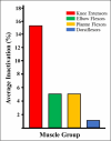
Figure 2.. Average inactivation by muscle group.
Relative to other muscle groups within the human body, the knee extensors (i.e., quadriceps femoris) have the largest inactivation percentage. This higher inactivation percentage suggests maximal, volitional recruitment/activation of the quadriceps is a challenge at baseline (i.e., before knee injury or surgery). %, percentage.
A group of graphs showing different types of functions Description automatically generated with medium confidence
Figure 3.. Length-tension properties of the quadriceps muscle group.
Previous work has outlined the sarcomere lengths of each quadriceps muscle throughout its functional operating range. The rectus femoris is the only quadriceps muscle that operates within the ascending limb of the length-tension curve. At any degree of knee flexion, the single-joint quadriceps muscles (i.e., vastus medialis, intermedius and lateralis) have sarcomere lengths that are working within the plateau to descending limb of the length-tension curve. %, percentage; μm, micrometers; RFE, residual force enhancement.
A graph of a person's knee flexion angle Description automatically generated
Figure 4.. The internal moment arm curves of the quadriceps muscle group.
The internal moment arm curve for the single-joint quadriceps muscles slightly increases and peaks from 0-30 degrees of knee flexion, then gradually decreases from 30-100 degrees. Similarly, the internal moment arm curve of rectus femoris peaks between 20-30 degrees of knee flexion but has a slightly steeper moment arm curve. Deg, degrees; mm, millimeters; red shading, range of knee flexion associated with peak internal knee extension moment.
A graph of a curved line Description automatically generated
Figure 5.. Force-ratio between the quadriceps and patellar tendons.
As the knee moves into deeper flexion, force transmission within the quadriceps tendon increases relative to the patellar tendon (a); this change in force-ratio is due to a decreasing quadriceps tendon internal moment relative to the patellar tendon (see Figure 6) with a concurrent increase in passive tension within the quadriceps muscle group (b). F, force; PT, patellar tendon; QT, quadriceps tendon; RF, rectus femoris; VL, vastus lateralis; VM, vastus medialis.
A collage of images of a human body Description automatically generated
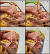
Figure 6.. Internal moment arms of the quadriceps and patellar tendons.
Between 0-30 degrees of knee flexion, the internal moment arm of the quadriceps tendon is larger than the patellar tendon (a, b). The patellar tendon has a larger internal moment arm than the quadriceps tendon in deeper levels of knee flexion (c, d). Blue line, patellar tendon internal moment arm; blue shading, patellar tendon; dotted black lines; resultant intratendinous force-vector from a quadriceps contraction; green line, quadriceps tendon internal moment arm; green shading, quadriceps tendon; red circle; estimated center of joint rotation; yellow shading, patella.
Figure 7.
Figure 7.. Depiction of the Knee Extensor Force-Velocity Relationship.
The force-velocity relationship for the quadriceps produces maximal concentric knee extension force/torque at slow contraction velocities (i.e., maximal effort pushing/overcoming isometric contractions or concentric contractions at < 60 degrees/second). Partially due to residual force enhancement during eccentric contractions (i.e., active muscle lengthening at maximal effort), maximal knee extension force/torque-output will be higher during eccentric than concentric contractions. %, percentage; 1-RM(con), 1-repitition maximum during a concentric contraction; deg, degrees; RFE, residual force enhancement; sec, second.
Figure 8.
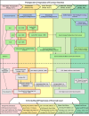
Figure 8.. Overview of exercise selection for quadriceps training after anterior cruciate ligament reconstruction (ACLR).
After anterior cruciate ligament reconstruction, quadriceps training should begin with quadriceps setting and be developed onto both open and closed-kinetic-chain load progressions. Strength training load progressions should be primarily advanced through the progression of external load, and exercises that load the quadriceps close to volitional fatigue/task failure at relatively long muscle lengths should be prescribed to enhance muscular hypertrophy. Lastly, a specific load progression to enhance quadriceps rate-of-force development should be prescribed throughout rehabilitation. 1-RM, concentric one-repetition maximum of the surgical limb; AD, assistive device; BFR, blood flow restriction; quad set, quadriceps setting exercise; black arrows, exercise progression pathway; blue boxes, exercise progression for quadriceps rate-of-force development; boxes with red glow, exercise with blood flow restriction; CKC, closed-kinetic-chain; green boxes, open-kinetic-chain exercise selection; grey boxes, closed-kinetic-chain exercise selection; LAQ, long-arc-quad exercise; MAX, maximal; NPRS, numeric pain rating scale; OKC, open-kinetic-chain; RFD, rate-of-force development; SAQ, short-arc-quad exercise; SCC, stretch-shortening cycle; SLR, straight leg raise exercise; SUBMAX, submaximal; TKE, terminal knee extension.
A collage of a person's legs Description automatically generated
Figure 9.. Exercise selection for the immediate postoperative phase.
Positioning the knee in 20-45 degrees of knee flexion prior to quadriceps setting will improve global quadriceps length-tension and its internal moment arm(s) (a). Slight femoral external rotation with active ankle dorsiflexion should be maintained throughout the straight leg raise exercise, as this position may maximize force output from the quadriceps (b). The CKC quadriceps load-progression should be initiated in sitting with the terminal knee extension exercise (i.e., isotonic, terminal knee extension with elastic band resistance); this exercise can be utilized to preferentially activate the distal / single-joint quadriceps muscles (c, d). Black lines, approximate tibial and femoral bone markers; dotted red line, midline/neutral hip rotation within the surgical limb; red shading, depiction of a knee flexion angle of 20-45 degrees; white circle; estimated center of joint rotation.
A person in shorts and a blue shirt Description automatically generated
Figure 10.. Closed-Kinetic-Chain Yielding/Holding Isometric Load-Progression.
The closed-kinetic-chain (CKC) yielding/holding isometric load progression should start with the double-leg squat exercise in low levels of knee flexion (a, non-surgical limb posted). Exercise selection can be advanced to include the split-squat exercise (b), and a heel wedge can be utilized to increase external knee flexion moment/load on the quadriceps (c). Submaximal yielding/holding isometric contractions should be advanced onto maximal effort contractions through increasing the degree of knee flexion/external knee flexion moment (d) or the progression of external load. Maximal effort CKC machine or weightbearing exercises with upper body stabilization (c, d) are preferred to free-weight or compound exercises prescribed without external support. Asterisk, use of wall support to stabilize the body during exercise (c, d); black lines, approximate tibial and femoral bone markers; red arrows; approximate ground reaction force-vector; white arrows, progression of knee/quadriceps load through the advancement of exercise position (a-c); white circle; estimated center of joint rotation; yellow arrow, progression of knee/quadriceps load by advancing the degree of knee flexion/external knee flexion moment (d); yellow shading; depiction of knee/quadriceps-dominant loading with increasing external knee flexion moment (a-d).
A collage of a person on a machine Description automatically generated
Figure 11.. Open and closed-kinetic-chain eccentric quasi-isometric quadriceps contractions.
Eccentric quasi-isometric (EQI) quadriceps resistance training can be completed on a leg press with a decline ankle-wedge (a-c) or knee extension machine (d-f) by (1) performing a concentric contraction to externally load the surgical knee/limb (a, d), (2) performing a yielding/holding isometric contraction to the point of volitional fatigue/failure (b, e), and (3) continuing to maximally resist the ensuing eccentric contraction throughout the prescribed range of knee motion. Black arrows, contraction direction; black lines, approximate tibial and femoral bone markers; green shading, concentric contractions; red shading, ensuing eccentric contractions as part of the EQI contraction; white asterisk, heel wedge; white circle, estimated center of joint rotation; yellow shading, depiction of large external knee flexion moment during yielding/holding isometric contractions
Figure 12.
Figure 12.. Preferred exercise technique for eccentric-specific resistance training.
Eccentric-specific open and closed-kinetic-chain quadriceps resistance training should be progressed onto load-intensities > 100% of the surgical limb’s concentric 1-repitition maximum. Eccentric-specific quadriceps training can be completed on a leg press with a decline ankle-wedge (a-d) or knee extension machine (e-h) by (1) performing a concentric contraction with both limbs from the machine’s bottom position (a, e) to the top position (b, f), (2) removing/moving the non-surgical limb to place all load on the surgical limb (c, g), and (3) eccentrically lowering the load through the desired range of knee motion. Green shading, concentric phase of exercise; red shading, eccentric phase of exercise; white asterisk, heel wedge; yellow asterisk; movement of the non-surgical limb throughout exercise.
A collage of a person lifting a weight Description automatically generated
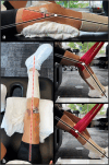
Figure 13.. Example of velocity-based training with a linear positioning transducer/accelerometer.
Velocity-based training methods can be utilized after anterior cruciate ligament reconstruction to improve quadriceps motor recruitment and rate-of-force development (a-d). To best stimulate improvements in peak quadriceps contraction-velocity/knee power, emphasis should be placed on the importance of a maximal effort/intent on each repetition. Black lines, approximate tibial and femoral bone markers; black circle, feedback monitor from linear positioning transducer/accelerometer; blue lines, ripcord connecting the exercise bar to the linear positioning transducer/accelerometer; red arrows; approximate ground reaction force-vector; white circles; estimated center of joint rotation; yellow shading; depiction of external knee flexion moment.

References

    1. Anterior cruciate ligament injury risk in sport: A systematic review and meta-analysis of injury incidence by sex and sport classification. Montalvo A. M., Schneider D. K., Webster K. E.., et al. 2019J Athl Train. 54(5):472–482. doi: 10.4085/1062-6050-407-16. https://doi.org/10.4085/1062-6050-407-16 - DOI - DOI - PMC - PubMed
    1. Anatomical versus non-anatomical single bundle anterior cruciate ligament reconstruction: A cadaveric study of comparison of knee stability. Lim H.-C., Yoon Y.-C., Wang J.-H., Bae J.-H. 2012Clin Orthop Surg. 4(4):249. doi: 10.4055/cios.2012.4.4.249. https://doi.org/10.4055/cios.2012.4.4.249 - DOI - DOI - PMC - PubMed
    1. Anatomical anterior cruciate ligament reconstruction (ACLR) results in fewer rates of atraumatic graft rupture, and higher rates of rotatory knee stability: a meta-analysis. Eliya Y., Nawar K., Rothrauff B. B., Lesniak B. P., Musahl V., de SA D. 2020Journal of ISAKOS. 5:359–370. doi: 10.1136/jisakos-2020-000476. https://doi.org/10.1136/jisakos-2020-000476 - DOI - DOI
    1. Treatment after anterior cruciate ligament injury: Panther Symposium ACL Treatment Consensus Group. Diermeier T., Rothrauff B. B., Engebretsen L.., et al. 2020Orthop J Sports Med. 8:232596712093109. doi: 10.1177/2325967120931097. https://doi.org/10.1177/2325967120931097 - DOI - DOI - PMC - PubMed
    1. Why bone-patella tendon-bone grafts should still be considered the gold standard for anterior cruciate ligament reconstruction. Carmichael J. R., Cross M. J. 2009Br J Sports Med. 43:323–325. doi: 10.1136/bjsm.2009.058024. https://doi.org/10.1136/bjsm.2009.058024 - DOI - DOI - PubMed

LinkOut - more resources