. 2024 Jul 3;34(27):2312038.
doi: 10.1002/adfm.202312038. Epub 2024 Mar 5.

Oxidized mRNA Lipid Nanoparticles for In Situ Chimeric Antigen Receptor Monocyte Engineering

Affiliations

Oxidized mRNA Lipid Nanoparticles for In Situ Chimeric Antigen Receptor Monocyte Engineering

Alvin J Mukalel et al. Adv Funct Mater. .

Abstract

Chimeric antigen receptor (CAR) monocyte and macrophage therapies are promising solid tumor immunotherapies that can overcome the challenges facing conventional CAR T cell therapy. mRNA lipid nanoparticles (mRNA-LNPs) offer a viable platform for in situ engineering of CAR monocytes with transient and tunable CAR expression to reduce off-tumor toxicity and streamline cell manufacturing. However, identifying LNPs with monocyte tropism and intracellular delivery potency is difficult using traditional screening techniques. Here, ionizable lipid design and high-throughput in vivo screening are utilized to identify a new class of oxidized LNPs with innate tropism and mRNA delivery to monocytes. A library of oxidized (oLNPs) and unoxidized LNPs (uLNPs) is synthesized to evaluate mRNA delivery to immune cells. oLNPs demonstrate notable differences in morphology, ionization energy, and pKa, therefore enhancing delivery to human macrophages, but not T cells. Subsequently, in vivo library screening with DNA barcodes identifies an oLNP formulation, C14-O2, with innate tropism to monocytes. In a proof-of-concept study, the C14-O2 LNP is used to engineer functional CD19-CAR monocytes in situ for robust B cell aplasia (45%) in healthy mice. This work highlights the utility of oxidized LNPs as a promising platform for engineering CAR macrophages/monocytes for solid tumor CAR monocyte therapy.


Figures

Figure 1.
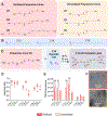
Figure 1.. Oxidized mRNA lipid nanoparticles for in situ CAR monocyte engineering.
(A) Ionizable lipid design and screening were used to identify oxidized ionizable lipids, with an internal ether chemical motif, that enhanced mRNA delivery to macrophages. (B) High throughput in vivo screening via DNA barcoding combined with mRNA validation studies were used to identify mRNA LNPs capable of engineering CAR monocytes directly in situ for solid tumor immunotherapy.
Figure 2.
Figure 2.. Oxidized and Unoxidized ionizable lipids formulate into LNPs with different ionization and morphological properties.
(A) Oxidized or unoxidized polyamine cores were designed to test the role of ether spacers for mRNA delivery. (B) Epoxide-terminated alkyl tails had a 12, 14, or 16 carbon alkyl chain. (C) Epoxide tails from (B) and polyamine cores from (A) were combined in a 7:1 molar ratio via SN2 reaction to synthesize a library of 18 ionizable lipids. (D) pKa characterization and ionization energy (E) measurements for 18 ionizable lipid library was measured by a 2-(p-toluidine)-6-naphthalenesulfonic acid (TNS) fluorescence assay. (F) Representative cryo-TEM images of LNPs formulated with a C14-O2 ionizable lipid and its equivalent unoxidized ionizable lipid, C14-U2. TNS fluorescence intensities were compared using a 2-way ANOVA with a Holm-Sidak correction. * p < .05, ** p < .01, *** p < .005, **** p < .001
Figure 3.
Figure 3.. oLNPs deliver luciferase mRNA to phagocytes with higher potency than uLNP counterparts.
(A) THP-1 human macrophages were treated with luciferase mRNA LNPs at a dose of 200 ng per 60k cells. Luminescence was measured 24 h later. n = 5 biological replicates. (B) Jurkat human T cells were treated with luciferase mRNA LNPs at a dose of 50 ng per 60k cells. Luminescence was measured 24 h later. n = 5 biological replicates. (C) Donor-derived CD14+ primary human monocytes were differentiated into macrophages after 7d culture in granulocyte-macrophage colony stimulating factor- (GMCSF) supplemented media. Macrophages were treated luciferase mRNA LNPs at a dose of 200 ng per 60k cells. Luminescence was measured 24 h later. n = 3 independent donors. (D) THP-1 human macrophages were treated with luciferase mRNA LNPs at a dose of 200 ng per 60k cells; cell viability was evaluated with a CellTiterGlo assay at 24 h. Luminescence signal was normalized to untreated cells. (E) Jurkat human T cells were treated with luciferase mRNA LNPs at a dose of 50ng per 60k cells; cell viability was evaluated with a CellTiterGlo assay at 24 h. (F) Primary human macrophages were generated as in (B). And treated with luciferase mRNA LNPs at a dose of 200 ng per 60k cells; cell viability was evaluated with a CellTiterGlo assay at 24 h. n = 3 independent donors. Luminescence signal was normalized to untreated cells. All data is presented as the mean ± standard deviation and was analyzed using a 2-way ANOVA with a Holm-Sidak correction. * p < .05, ** p < .01, *** p < .005, **** p < .001
Figure 4.
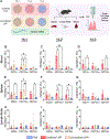
Figure 4.. High throughput screening via DNA barcoding identifies oxidized LNPs with tropism to immune cells (B cells, T cells, dendritic cells, macrophages).
(A) Overview of barcoding experiment: each LNP formulation encapsulated mCherry mRNA and a unique 61 nt DNA barcode, LNPs were pooled and injected to C57BL/6 mice via the tail vein, FACS isolated immune cell populations, and NGS identified LNPs with tropism to immune cell types. (B) B cells (CD19+), dendritic cells (CD83+), macrophages (CD11b+) and T cells (CD3+) were isolated from the blood, lymph nodes, and spleen; barcode accumulation in cells was normalized to an uninjected pool of the barcoded LNP library. Each LNP formulation is represented as a column. n = 5 mice. (C) Volcano plot enrichment diagrams comparing statistical significance of enrichment or depletion of individual barcodes relative to the rest of the pool. (D) Scoring of barcoded library used to identify lead candidate LNPs for functional mRNA validation.
Figure 5.
Figure 5.. In vivo evaluation of mCherry mRNA LNPs identifies oxidized C14-O2 LNP with potent and selective delivery to monocytes.
After DNA barcoding, 6 LNPs were evaluated for their ability to deliver mCherry mRNA in vivo. (A) Overview of in vivo validation screen with mCherry mRNA. C57BL/6 mice were injected via the tail vein at a dose of 1 mg kg−1, organs were dissected 12 h post injection. Immune cells were isolated from the spleen, lymph node and blood and mCherry expression was evaluated on a single cell level using flow cytometry. (B-J) Transfection rates for each oLNP and uLNP pair in immune cells isolated from the blood (B-D), spleen (E-G), and inguinal lymph nodes (H-J). n = 4 mice. All data is presented as the mean ± standard deviation and was analyzed using a 2-way ANOVA with Holm-Sidak correction. * p < .05, ** p < .01
Figure 6.
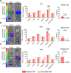
Figure 6.. Whole body biodistribution of top LNPs indicates oxidized LNPs preferentially deliver luciferase mRNA to the spleen.
(A) Representative images (left), organ luminescence quantification (middle) and quantified ratio of spleen to liver luminescence (right) for the C16–1 LNPs (A), C14–2 LNPs (B), and C14–3 LNPs (C). C57BL/6 mice were injected via the tail vein at a dose of 5 ug mRNA per mouse. After 6 h, mice were sacrificed, and major organs were harvested and analyzed for luciferase expression using IVIS imaging. n = 3–4 mice per group. Organs in IVIS images from top to bottom: heart, lungs, liver, kidneys, spleen, lymph nodes. Luminescence signal is normalized to PBS treated mice. All data is presented as the mean ± standard deviation. Organ luminescence was compared between oLNPs and uLNPs using a 2-way ANOVA with Holm-Sidak correction. * p < .05, ** p < .01, *** p < .005, **** p < .001. Spleen:liver ratios were compared between oLNPs and uLNPs using an unpaired t-test.
Figure 7.
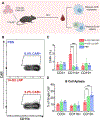
Figure 7.. Oxidized C14-O2 LNP engineers functional CD19-CAR monocytes directly in situ and induces B cell aplasia.
B) Representative flow plots and quantification (C) of CD19-CAR expression in blood monocytes isolated from mice treated with PBS or C14-O2 LNP encapsulating CD19-CAR mRNA. D) Healthy mice receiving CAR LNPs have significantly reduced CD19+ blood B cells compared to mice receiving luciferase LNPs or PBS. C57BL/6 mice were injected via the tail vein at a dose of 1 mg kg−1 and blood was collected 12 h post injection. n = 4 mice per group. All data is presented as the mean ± standard deviation and was analyzed using a 2-way ANOVA with Holm-Sidak correction. * p < .05, ** p < .01, *** p < .005, **** p < .001

References

    1. Schuster SJ et al. Chimeric Antigen Receptor T Cells in Refractory B-Cell Lymphomas. New England Journal of Medicine 377, 2545–2554 (2017). - PMC - PubMed
    1. Park JH et al. Long-Term Follow-up of CD19 CAR Therapy in Acute Lymphoblastic Leukemia. New England Journal of Medicine 378, 449–459 (2018). - PMC - PubMed
    1. Neelapu SS et al. Chimeric antigen receptor T-cell therapy — assessment and management of toxicities. Nat Rev Clin Oncol 15, 47–62 (2018). - PMC - PubMed
    1. Maude SL et al. Tisagenlecleucel in Children and Young Adults with B-Cell Lymphoblastic Leukemia. New England Journal of Medicine 378, 439–448 (2018). - PMC - PubMed
    1. CAR T Cells: Engineering Immune Cells to Treat Cancer - NCI. https://www.cancer.gov/about-cancer/treatment/research/car-t-cells (2013).

LinkOut - more resources