Co-Existing Ectopic Cortisol-Producing Adenoma and Retroperitoneal Schwannoma, a Rare Case Report
- PMID: 39629070
- PMCID: PMC11612559
- DOI: 10.2147/DMSO.S487334
Co-Existing Ectopic Cortisol-Producing Adenoma and Retroperitoneal Schwannoma, a Rare Case Report
Abstract
Background: Ectopic cortisol-producing adrenocortical adenoma (ECPA) is extremely rare, with only a few cases reported. Retroperitoneal schwannoma is also uncommon, accounting for only 0.7-5% of all schwannomas. It is peculiar to have both conditions at the same time, and it is intriguing to explore their possible connection. Herein, we present a case of both ECPA and retroperitoneal schwannoma and provide our conjectures regarding their co-occurrence.
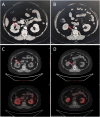
Case presentation: A 38-year-old female presented with a two-year history of facial and lower limb edema, as well as chest tightness and palpitations for the past four months. Physical examination revealed hypertension, a high body mass index (BMI), moon face, thick neck and back fat, abdominal obesity, and purple striae on the abdomen. Laboratory tests indicated an persistent increased cortisol level and suppression of adrenocorticotropic hormone (ACTH). Adrenal-enhanced computed tomography (CT) scan showed that both adrenal glands appeared normal without evident adenomas or hyperplasia. However, the scan revealed two lesions located in the right renal hilum and retroperitoneal area positioned anteriorly to the lower margin of the lumbar 2 pyramid. Further imaging using 68Ga-DOTATATE PET/CT revealed concentrated radiotracer uptake in the tumor at the right renal hilum, indicating it may be responsible for the patient's Cushing's symptoms. After laparoscopic resection of these masses, clinical symptoms improved significantly. Postoperative pathology confirmed the right renal hilum one lesion as an ECPA while identifying another lesion as a schwannoma.
Conclusion: Our literature reported a case with the diagnosis of both ECPA in the right renal hilum and retroperitoneal schwannoma. 68Ga-DOTATATE PET/CT imaging can provide functional and locational information on tumors, enabling a comprehensive examination of the entire body to identify lesions that require appropriate treatment.
Keywords: Cushing syndrome; case report; cortisol-producing adrenocortical adenomas; ectopic adrenocortical adenoma; schwannoma.
© 2024 Shi et al.
Conflict of interest statement
The authors declare that they have no competing interests.
Figures


References
Publication types
LinkOut - more resources
Full Text Sources
Miscellaneous

