. 2024 Oct 28;35(4):102374.
doi: 10.1016/j.omtn.2024.102374. eCollection 2024 Dec 10.

Exploring non-coding variants and evaluation of antisense oligonucleotides for splicing redirection in Usher syndrome

Affiliations

Exploring non-coding variants and evaluation of antisense oligonucleotides for splicing redirection in Usher syndrome

Belén García-Bohórquez et al. Mol Ther Nucleic Acids. .

Abstract

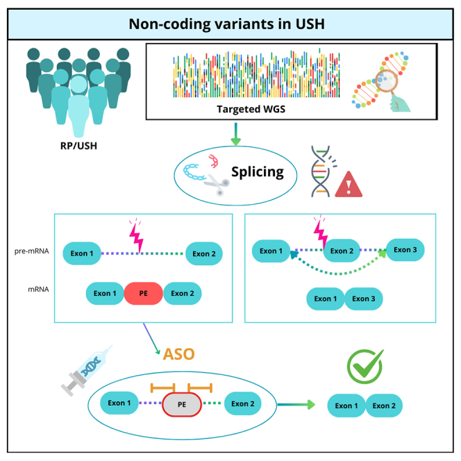
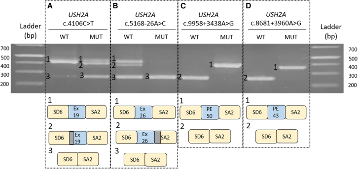
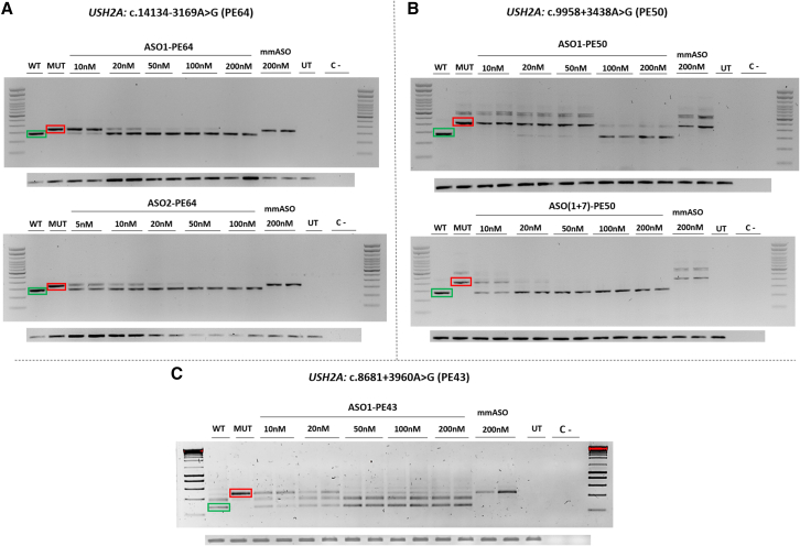
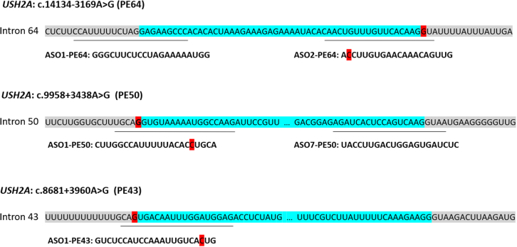
Exploring non-coding regions is increasingly gaining importance in the diagnosis of inherited retinal dystrophies. Deep-intronic variants causing aberrant splicing have been identified, prompting the development of antisense oligonucleotides (ASOs) to modulate splicing. We performed a screening of five previously described USH2A deep-intronic variants among USH2A monoallelic patients with Usher syndrome (USH) or isolated retinitis pigmentosa. Sequencing of entire USH2A or USH genes was then conducted in unresolved or newly monoallelic cases. The splicing impact of identified variants was assessed using minigene assays, and ASOs were designed to correct splicing. The screening allowed to diagnose 30.95% of the studied patients. The sequencing of USH genes revealed 16 new variants predicted to affect splicing, with four confirmed to affect splicing through minigene assays. Two of them were unreported deep-intronic variants and predicted to include a pseudoexon in the pre-mRNA, and the other two could alter a regulatory cis-element. ASOs designed for three USH2A deep-intronic variants successfully redirected splicing in vitro. Our study demonstrates the improvement in genetic characterization of IRDs when analyzing non-coding regions, highlighting that deep-intronic variants significantly contribute to USH2A pathogenicity. Furthermore, successful splicing modulation through ASOs highlights their therapeutic potential for patients carrying deep-intronic variants.

Keywords: MT: Oligonucleotides: Therapies and Applications; USH2A; Usher syndrome; antisense oligonucleotides; deep-intronic; minigene; non-coding regions; pseudoexon; splicing.


Conflict of interest statement

The authors declare no competing interests.

Figures

None
Graphical abstract
Figure 1
Figure 1
Analyses of potential splicing modulating variants in USH2A using minigene splice assays SD6 and SA2 are the constitutive exons of pSPL3 plasmid and in blue boxes exons (ex) and pseudoexons (PEs) are depicted. The first three variants present a(n) (partial) exon-skipping effect, while the last two have included a PE in their sequence. Boxes in gray represent regions that have not been included in the sequence as an alternative splicing has occurred.
Figure 2
Figure 2
ASO-induced redirection of aberrant splicing caused by deep-intronic variants Each section represents WT (green) and MUT (red) fragments and the different treatment dose. The lower panels in every section are the GAPDH loading control in each experiment. For PE 64 (A), two different ASO were tested separately. For PE50 (B), ASO7-PE50 was tested together with ASO1-PE50 due to the lower efficiency it showed individually and the concentrations shown for ASO(1+7)-PE50 refer to the final concentration of both ASO. For PE43 (C), only ASO1-PE43 was tested as the sequence did not agree with all the requirements. Differences between wild-type and mutant fragments of PE50 and PE43 constructs in Figure 1 compared with this figure can be observed due to the use of different cloning splice vector for each experiment. WT, wild type; MUT, mutant; UT, untreated; C–, negative control.
Figure 3
Figure 3
Position of the designed ASOs with respect to the different PEs Sequences in gray represent the intronic region, whereas the blue nucleotides correspond to the PEs. Variants are depicted in red. The lines below the different intron indicate the specific sequence for each ASO, which are also empathized right under them. PE50 and PE43 are disrupted by dots to make all sequences equal in length.

References

    1. Heath Jeffery R.C., Mukhtar S.A., McAllister I.L., Morgan W.H., Mackey D.A., Chen F.K. Inherited retinal diseases are the most common cause of blindness in the working-age population in Australia. Ophthalmic Genet. 2021;42:431–439. doi: 10.1080/13816810.2021.1913610. - DOI - PMC - PubMed
    1. Verbakel S.K., van Huet R.A.C., Boon C.J.F., den Hollander A.I., Collin R.W.J., Klaver C.C.W., Hoyng C.B., Roepman R., Klevering B.J. Non-syndromic retinitis pigmentosa. Prog. Retin. Eye Res. 2018;66:157–186. doi: 10.1016/j.preteyeres.2018.03.005. - DOI - PubMed
    1. Tatour Y., Ben-Yosef T. Syndromic Inherited Retinal Diseases: Genetic, Clinical and Diagnostic Aspects. Diagnostics. 2020;10:779. doi: 10.3390/diagnostics10100779. - DOI - PMC - PubMed
    1. Espinós C., Millán J.M., Beneyto M., Nájera C. Epidemiology of Usher Syndrome in Valencia and Spain. Public Health Genomics. 1998;1:223–228. doi: 10.1159/000016167. - DOI - PubMed
    1. Kimberling W.J., Hildebrand M.S., Shearer A.E., Jensen M.L., Halder J.A., Trzupek K., Cohn E.S., Weleber R.G., Stone E.M., Smith R.J.H. Frequency of Usher syndrome in two pediatric populations: Implications for genetic screening of deaf and hard of hearing children. Genet. Med. 2010;12:512–516. doi: 10.1097/GIM.0b013e3181e5afb8. - DOI - PMC - PubMed

LinkOut - more resources