. 2024 Oct 21;14(18):7023-7041.
doi: 10.7150/thno.97662. eCollection 2024.

TRIM4 enhances small-molecule-induced neddylated-degradation of CORO1A for triple negative breast cancer therapy

Affiliations

TRIM4 enhances small-molecule-induced neddylated-degradation of CORO1A for triple negative breast cancer therapy

Wen-Jie Gu et al. Theranostics. .

Abstract

Background: As a critical member of the Coronin family, Coronin 1A (CORO1A) plays a crucial role in the progression of triple-negative breast cancer (TNBC). However, CORO1A is typically considered "undruggable" due to its smooth surface and complex protein-protein interactions (PPIs). Molecular glues have emerged as one of the most effective strategies to rapidly degrade such "undruggable" targets. Neddylation, an emerging approach, has shown promise in targeting pathogenic proteins for degradation through the NEDD8 pathway, making the degradation of CORO1A an attractive pharmacological strategy. Methods: A phenotypic drug screening strategy coupled with multi-omics approaches was utilized to rapidly identify a molecular glue degrader for CORO1A and to uncover the associated mechanisms. The Omics and Text-based Target Enrichment and Ranking (OTTER) tools, co-immunoprecipitation (Co-IP) assay, mass spectrometry, and the separation of phases-based protein interaction reporter (SPPIER) method were employed to explore the interaction between Aurovertin B (AB) and CORO1A via TRIM4. The pharmacological effects of AB were assessed using TNBC patient-derived organoids (PDOs) and 3D bioprinting models. Results: We identified AB as a previously undisclosed molecular glue that significantly promotes the neddylation and proteasomal degradation of CORO1A via TRIM4, an atypical E3 ligase. Notably, the degradation of CORO1A markedly inhibited various cellular processes and exerted robust antitumor effects in TNBC PDOs and 3D bioprinting models. Conclusions: Our findings underscore the critical role of CORO1A in TNBC and lay a crucial foundation for the development of innovative drugs based on molecular glue technology.

Keywords: Aurovertin B; CORO1A; Molecular glues; Neddylation; Triple-negative breast cancer.


Conflict of interest statement

Competing Interests: The authors have declared that no competing interest exists.

Figures

Figure 1
Figure 1
CORO1A overexpression correlated with poor clinical outcomes in TNBC patients. (A) Pan-cancer analysis of CORO1A using the GEPIA database. CORO1A expression was significantly increased in BC patients using the GSE21422 database (B). (C) CORO1A expression was significantly increased in patients with TNBC (GSE27447 database). (D) CORO1A overexpression was observed in TNBC patients using tissue chip staining. The H-score of CORO1A was showed in (G). Representative images of colony formation were shown in MDA-MB-231 cells with shCORO1A (E) and in MDA-MB-468 cells with siCORO1A (F). The analysis was showed in (H) compared to the Scramble group. (I) The expression of CORO1A in different subtypes of BC was higher than that in normal tissues, especially in TNBC. (J) A high level of CORO1A was correlated with poor prognosis in TNBC patients. (K) Tumorigenicity experiments showed that the MDA-MB-231 cells with shCORO1A failed to promote tumor growth. The EMT (L) and TGFβ pathway (M) scores correlated highly with CORO1A across BC tumors. Data were presented as mean ± SD, ***P ˂ 0.001.
Figure 2
Figure 2
AB exerted anti-TNBC effects by directly binding to CORO1A. (A) The pattern diagram of virtual screening and the chemical structure of AB. (B) Cell viability of MDA-MB-231 and MDA-MB-468 cells after AB treatment for 24 h compared to group with no drug treatment. (C) Transwell assay showed that AB inhibited cell migration compared with no drug treatment. (D) A flow cytometry assay showed AB-induced cell apoptosis compared with no drug treatment. (E) Representative images of Live/Dead staining of cells with 3D bioprinting. (F) CETSA assays verified the destabilization of CORO1A in MDA-MB-231 cells. (G) DARTS assays confirmed the destabilization of CORO1A during proteolysis in MDA-MB-231 cells. (H) Analysis of (F). (I) Analysis of (G). (J) MST analysis of the binding of GFP-tagged CORO1A with AB. (K) Molecular docking results of AB binding to CORO1A. (L) MST analysis of the binding of GFP-tagged mutant CORO1A with AB. Data were presented as mean ± SD, *P ˂ 0.05, **P ˂ 0.01, ***P ˂ 0.001 versus the control group.
Figure 3
Figure 3
CORO1A underwent neddylation-dependent degradation induced by AB. (A) AB inhibited the protein expression of CORO1A in MDA-MB-231 and MDA-MB-468 cells. The analysis was showed in (B) and (C). (D) The representative microscopic photographs of the CORO1A expression in MDA-MB-231 and MDA-MB-468 cells after AB (0.2 μM) treatment for 24 h. The analysis was shown in (E) and (F). (G) AB accelerated the degradation of CORO1A in MDA-MB-231 cells. The analysis was showed in (H). (I) MG132 (0.5 μM) rescued the degradation of CORO1A protein induced by AB (0.2 μM) in MDA-MB-231 cells. The analysis was shown in (J). (K) The pattern diagram of OTTER analysis. (L) The pattern diagram of transcriptome sequencing. (M) The top 20 ubiquitin-related proteins were obtained using OTTER enrichment analysis. (N) MLN4924 reversed the degradation of CORO1A. (O) Analysis of (N). (P) MLN4924 attenuated the inhibitory effect of AB on MDA-MB-231 cells. (Q) WS383 attenuated the inhibitory effect of AB on MDA-MB-231 cells. Data were presented as mean ± SD, *P ˂ 0.05, ***P ˂ 0.001 versus the control group.
Figure 4
Figure 4
Identification of AB as a molecular glue degrader for CORO1A. (A) Co-IP assay using non-denaturing conditions confirmed the neddylation of CORO1A. (B) The pattern diagram of immunoprecipitation (IP) for LC-MS/MS analysis. (C) The proteins that interacted with CORO1A were identified by combining Co-IP and mass spectrometry assay. (D) TRIM4 changed remarkably upon AB treatment. (E) Co-IP assay in 293T cells confirmed the increased binding of TRIM4 to CORO1A after AB treatment. (F) Analysis of the relative TRIM4 level binding with CORO1A after AB treatment in IP group. (G) Knockdown of TRIM4 attenuated the inhibitory effect of AB on MDA-MB-231 cells. (H) Molecular docking studies suggested that AB could bind to multiple sites of TRIM4 with a docking score of -8.26. (I) Molecular dynamics simulations showed that AB could stabilize the binding of CORO1A to TRIM4. (J) SPPIER assay visualized ternary complex formation. The analysis was conducted in (K). Data were presented as mean ± SD, *P ˂ 0.05, **P ˂ 0.01, ***P ˂ 0.001 versus the group of AB treatment.
Figure 5
Figure 5
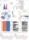
CORO1A degradation regulated TGFβ-Smads signaling and cell apoptosis. (A) The volcano plot represents differentially expressed genes. (B) KEGG enrichment analysis of quantitative proteomics. (C) Network of pathways that down-regulated with AB. (D) GSEA analysis showed a significantly decreased TGF-β signaling pathway induced by AB. (E) The heatmap analysis of down-regulated genes. (F) Western blot assay proved that AB downregulated TGFβ-Smads signaling. (G) Knockdown of CORO1A affected TGFβ-Smads signaling. (H) Analysis of (F). (I) Analysis of (G). Data were presented as mean ± SD, ***P ˂ 0.001 versus the control group.
Figure 6
Figure 6
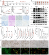
AB showed potent antitumor effects in TNBC-derived CDX models and PDO models. (A) Schematic diagram of in vivo study. (B) When mice were sacrificed, the tumors were photographed (n = 6 mice). (C) Tumor volume. (D) Body weight. (E) Tumor weight. (F) Western blot assay to detect the protein expression in tumor tissues. (G) IHC staining showed that AB induced increased necrosis and apoptosis, decreased proliferation, and decreased CORO1A expression. (H) Analysis of (G). (I) Analysis of (F). (J) AB inhibited TNBC PDOs formation with an IC50 value of 63.16 nM. (K) Representative images of Live/Dead staining of PDOs. Data were presented as mean ± SD, *P ˂ 0.05, **P ˂ 0.01, ***P ˂ 0.001 versus the control group.

References

    1. Sung H, Ferlay J, Siegel RL, Laversanne M, Soerjomataram I, Jemal A. et al. Global Cancer Statistics 2020: GLOBOCAN Estimates of Incidence and Mortality Worldwide for 36 Cancers in 185 Countries. Ca-Cancer J Clin. 2021;71:209–49. - PubMed
    1. Leon-Ferre RA, Goetz MP. Advances in systemic therapies for triple negative breast cancer. Bmj-Brit Med J. 2023;381:e071674. - PubMed
    1. Torres QS, Canha-Borges A, Oliveira MJ, Sarmento B, Castro F. Special Issue: Nanotherapeutics in Women's Health Emerging Nanotechnologies for Triple-Negative Breast Cancer Treatment. Small. 2023;20:e2300666. - PubMed
    1. Jaiswal A, Kaushik N, Choi EH, Kaushik NK. Functional impact of non-coding RNAs in high-grade breast carcinoma: Moving from resistance to clinical applications: A comprehensive review. Biochim Biophys Acta Rev Cancer. 2023;1878:188915. - PubMed
    1. Xavier CP, Eichinger L, Fernandez MP, Morgan RO, Clemen CS. Evolutionary and functional diversity of coronin proteins. Subcell Biochem. 2008;48:98–109. - PubMed

Publication types

MeSH terms