Review
. 2024 Oct 21;14(18):7111-7121.
doi: 10.7150/thno.102552. eCollection 2024.

Molecular imaging along the heart-kidney axis

Affiliations
Review

Molecular imaging along the heart-kidney axis

Konrad Klimek et al. Theranostics. .

Abstract

Cardiorenal syndrome (CRS) involves bidirectional crosstalk between the failing heart and the kidneys. Depending on the primum movens (primary cardiac or renal injury), systems-based interactions in the secondary affected organ may include pro-fibrotic signaling, overzealous inflammation, impaired nerve integrity or overactivity of specific renal transporters mediating glucose absorption. Those pathophysiological pillars can be investigated by molecular imaging using SPECT or PET agents. Targeted whole-body molecular imaging may allow for a) systems-based analysis along the heart-kidney axis, b) may provide prognostic information on longitudinal organ-based functional decline or c) may be used for guidance of reparative intervention based on peak activation identified on PET (paradigm of cardiorenal theranostics). We will discuss the current state of translational molecular imaging for CRS, along with future clinical aspects in the field.

Keywords: PET; cardiorenal; cardiorenal syndrome; heart-kidney axis; molecular imaging; organ-organ interaction; renocardiac; theranostics.


Conflict of interest statement

Competing Interests: R.A.W.: speaker honoraria from Novartis/AAA and PentixaPharm, advisory board work for Novartis/AAA and Bayer. All other authors declare no conflict of interest.

Figures

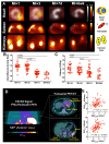
Figure 1
Figure 1
Overview of selected radiotracers used to decipher target expression along the heart-kidney axis. [123I]-meta-iodobenzylguanidine ([123I]mIBG), [11C]meta- hydroxyephedrine ([11C]HED), F18 N-[3-Bromo-4-(3-[18F]fluoro-propoxy)-benzyl]-guanidin ([18F]LMI1195), 2-Butyl-5-[11C]methoxymethyl-6-(1-oxopyridin-2-yl)-3-[[2-(1H-tetrazol-5-yl)biphenyl-4-yl]methyl]-3H-imidazo[4,5-b]pyridine ([11C]KR31173), 18F-labeled valsartan ([18F]FV45), 2-deoxy-2-[18F]fluoro-D-Glucose ([18F]FDG), 68Ga-labeled FAP inhibitor ([68Ga]FAPI04), lpha-methyl-4-deoxy-4-[18F]fluoro-D-glucopyranoside ([18F]Me-4FDG), 11C-methyl-D-glucoside ([11C]MDG). Created with biorender.com.
Figure 2
Figure 2
Cardiorenal C-X-C motif chemokine receptor 4 (CXCR4)-directed PET imaging using [68Ga]PentixaFor in mice (A-C) and patients (D, E) after myocardial infarction (MI). (A) [68Ga]PentixaFor revealed intense CXCR4 uptake early after MI (day [d] 1, day 3) in the infarct territory (short axis) and no relevant signal at later stages (day 7, week [wk] 6). Respective splenic and renal images (coronal axis) showed paralleled radiotracer decline over time. Quantitative evaluation provided by myocardial infarct to remote activity ratio (B) and renal [68Ga]PentixaFor signal (in %ID/g, C) confirmed visual findings with significant decline of in-vivo CXCR4 expression already at d7 relative to d1 post-MI. Maximum intensity projection (MIP) of [68Ga]PentixaFor in a patient early after MI revealed intense uptake in the infarct region, spleen and parenchyma of the kidneys (D). Peak standardized uptake values (SUVpeak) in the infarct region showed significant association with renal and splenic uptake (E), indicative for systemic immune response (as demonstrated by hematopoietic organ activation) and cardiorenal inflammatory interaction. Right ventricle (RV), left ventricle (LV), Spleen (S), L (Left), R (Right), K (Kidney). Adapted with permission from , copyright 2021 IvySpring.
Figure 3
Figure 3
Norepinephrine (NE) transporter (NET)-directed molecular using [123I]mIBG planar scintigraphy. In healthy patients (upper row, left), NE is cleared from the synaptic cleft in presynaptic nerve terminal once a firing impulse has arrived. In heart failure patients, neurotransmission is characterized by a compromised reuptake mechanism (upper row, right). Mimicking the physiological pathway of NE, the [123I]mIBG signal is then diminished in HF patients (lower row, right), while the radiotracer signal is intense in healthy individuals (lower row, left). Dotted circles indicate myocardium. SNS = sympathetic nervous system. Created with biorender.com.
Figure 4
Figure 4
SGLT-directed molecular imaging using [18F]Me-4FDG. (A) Relative to [18F]FDG (right panel), which interacts with GLUT, [18F]Me-4FDG is selectively taken up by SGLT (middle panel). (B) Renal [18F]Me-4FDG 10-20 min after i.v. injection (in healthy rats (left panels). Stable radiotracer accumulation was observed in controls, while in Phlorizin- and Canagliflozin-pretreated animals, uptake in the cortex was substantially decreased (along with high bladder activity). Cortex-to-pelvis count ratios (upper row, right) also displayed lower radiotracer accumulation over time relative to controls. Kidney and blood radioactivity (lower row, right) at 60 min after tracer i.v. showed increased renal activity only for controls, but not for pretreated animals, thereby indicating high specificity of [18F]Me-4FDG for deciphering SGLT activity in-vivo. Adapted and changed with permission from , copyright 2022 Sage. Published under Creative Commons Attribution 4.0 License (https://creativecommons.org/licenses/by/4.0/).
Figure 5
Figure 5
Paradigm of cardiorenal theranostics. In a manner similar to oncology, the concept of theranostics could be transferred to individuals with cardiorenal syndrome. Patients with increased radiotracer signal, e.g., after injection of inflammatory-directed [68Ga]PentixaFor, will receive targeted, anti-inflammatory therapy, thereby improving cardiorenal functional outcome. For patients with diminished PET signal in both target organs, other treatment options should be identified. As such, PET can then allow for a molecular image-piloted strategies to determine the most appropriate patient at the right time for the right treatment. Inconsistencies on cardiorenal uptake may be addressed by dose-depending therapeutic approaches, e.g., lower uptake may trigger lower dose of a cardiorenal protective agent.

References

    1. Rangaswami J, Bhalla V, Blair JEA, Chang TI, Costa S, Lentine KL. et al. Cardiorenal Syndrome: Classification, Pathophysiology, Diagnosis, and Treatment Strategies: A Scientific Statement From the American Heart Association. Circulation. 2019;139:e840–e78. - PubMed
    1. Heitzer E, Perakis S, Geigl JB, Speicher MR. The potential of liquid biopsies for the early detection of cancer. NPJ Precis Oncol. 2017;1:36. - PMC - PubMed
    1. Grenier N, Merville P, Combe C. Radiologic imaging of the renal parenchyma structure and function. Nat Rev Nephrol. 2016;12:348–59. - PubMed
    1. Buchanan CE, Mahmoud H, Cox EF, McCulloch T, Prestwich BL, Taal MW. et al. Quantitative assessment of renal structural and functional changes in chronic kidney disease using multi-parametric magnetic resonance imaging. Nephrol Dial Transplant. 2020;35:955–64. - PMC - PubMed
    1. Edwards NC, Moody WE, Chue CD, Ferro CJ, Townend JN, Steeds RP. Defining the natural history of uremic cardiomyopathy in chronic kidney disease: the role of cardiovascular magnetic resonance. JACC Cardiovasc Imaging. 2014;7:703–14. - PubMed

Publication types

MeSH terms

LinkOut - more resources