. 2024 Nov 19:11:1443368.
doi: 10.3389/fmed.2024.1443368. eCollection 2024.

Beneficial effects of non-invasive physical plasma on human periodontal ligament cells in vitro

Affiliations

Beneficial effects of non-invasive physical plasma on human periodontal ligament cells in vitro

Benedikt Eggers et al. Front Med (Lausanne). .

Abstract

Introduction: Periodontitis is a chronic inflammatory disease of the periodontium that can lead to the loss of affected teeth if left untreated. It is induced by a multifactorial process centered on microbial pathogens such as Fusobacterium nucleatum (F.n.). Non-invasive physical plasma (NIPP), a highly reactive gas, has become a focus of research, not only for its hemostatic, proliferation-enhancing and apoptotic properties, but also for its antimicrobial potential. The objective of this study was to examine the impact of NIPP on human periodontal ligament (PDL) cells that had been induced into a state of periodontal infection in vitro.

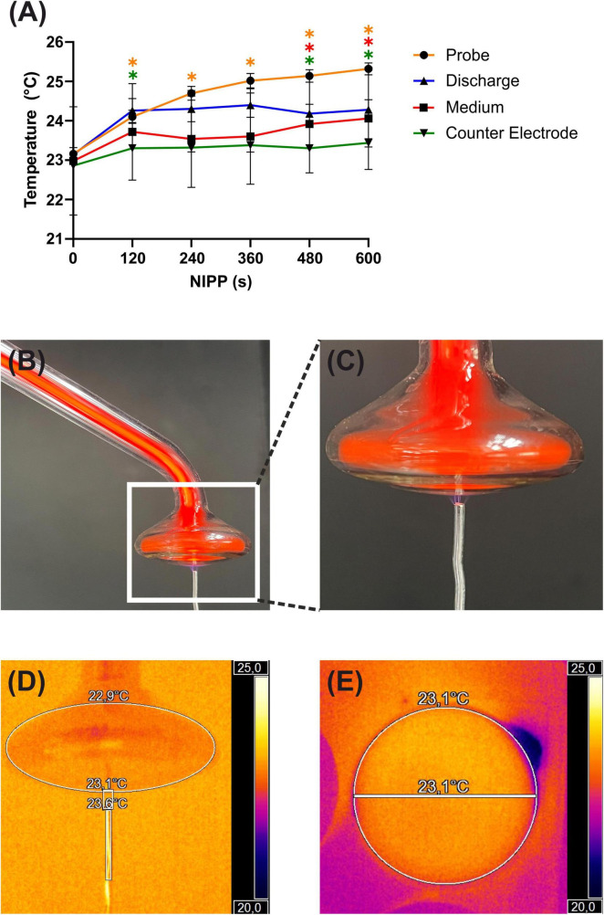
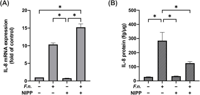
Methods: Initially, the solitary effect of NIPP was evaluated by measuring temperature and pH and analyzing reactive oxygen species (ROS). Additionally, DAPI and phalloidin staining were employed to investigate possible cytotoxic effects. The cells were pre-incubated with F.n. and treated with NIPP after 24 hours. Interleukin (IL)-6 and IL-8 were analyzed at mRNA and protein levels, respectively, by real-time PCR and ELISA.

Results: NIPP alone had no significant effect on PDL cells. However, the F.n.-induced upregulation of IL-6 and IL-8 was counteracted by NIPP.

Discussion: Thus, the utilization of NIPP may be regarded as a promising therapeutic strategy for the treatment of periodontal diseases.

Keywords: antimicrobial; cold plasma; human periodontal ligament cells; non-invasive physical plasma; non-thermal plasma; periodontology.


Conflict of interest statement

The authors declare that the research was conducted in the absence of any commercial or financial relationships that could be construed as a potential conflict of interest.

Figures

FIGURE 1
FIGURE 1
Basic characterization of NIPP during treatment: (A) temperature of the NIPP device and treated objects, n = 5. * Statistical significance compared to NIPP 0 s (p < 0.05). (B) Plasma ONE with probe PS30 and counter electrode in discharge mode. (C) Magnification of the probe tip. (D) Thermal image of the probe tip using the Optris PI 400i infrared camera with Optri PIX Connect analysis software (Optris, Berlin, Germany). (E) Thermal image of the bottom view of the probe tip.
FIGURE 2
FIGURE 2
Basic characterization of NIPP during treatment of medium: (A) pH of medium, n = 5 (B) O3 concentration in medium, n = 2 (C) H2O2 concentration in medium, n = 6 (D) NO3 concentration in medium, n = 5 (E) NO2 concentration in medium, n = 5. *Statistical significance compared to control (p < 0.05).
FIGURE 3
FIGURE 3
Immunofluorescence staining of ROS production in PDL cells, 10 min after NIPP treatment. Cells were treated for 0 s (A), 30 s (B), 120 s (C), 180 s (D). The scale bar represents 100 μm. ROS fluorescence was quantified using a fluorescent reader (E). n = 4. *Statistical significance (p < 0.05).
FIGURE 4
FIGURE 4
Immunofluorescence staining of periodontal ligament (PDL) cells with DAPI (blue staining of the nucleus) and phalloidin (green staining of the cytoskeleton) that were left untreated (A) or treated with NIPP for 30 s at 24 h (B). NIPP treatment showed no cytotoxic effects. The scale bar represents 100 μm.
FIGURE 5
FIGURE 5
IL-6 and IL-8 levels in PDL cells after pre-incubation with inactivated F.n. at different concentrations (OD = 0.025, 0.05, 0.01). (A) IL-6 mRNA expression at 24 h, n = 9 (B) IL-6 protein level at 48 h, n = 12 (C) IL-8 mRNA expression at 24 h, n = 9 (D) IL-8 protein level at 48 h, n = 12. *Statistical significance (p < 0.05).
FIGURE 6
FIGURE 6
IL-6 regulation in PDL cells after 24 h pre-incubation with inactivated F.n. at OD 0.025 (+) and after 30 s of NIPP treatment (+). (A) IL-6 mRNA expression at 24 h, n = 6 (B) IL-6 protein level at 48 h, n = 12. * Statistical significance (p < 0.05).
FIGURE 7
FIGURE 7
IL-8 regulation in PDL cells after 24 h pre-incubation with inactivated F.n. at OD 0.025 (+) and after 30 s of NIPP treatment (+). (A) IL-8 mRNA expression at 24 h, n = 6 (B) IL-8 protein level at 48 h, n = 12. * Statistical significance (p < 0.05).

Similar articles

References

    1. Pihlstrom B, Michalowicz B, Johnson N. Periodontal diseases. Lancet. (2005) 366:1809–20. - PubMed
    1. Trindade D, Carvalho R, Machado V, Chambrone L, Mendes J, Botelho J. Prevalence of periodontitis in dentate people between 2011 and 2020: A systematic review and meta-analysis of epidemiological studies. J Clin Periodontol. (2023) 50:604–26. 10.1111/jcpe.13769 - DOI - PubMed
    1. Deschner J, Schröder A, Weber M, Galler K, Proff P, Kirschneck C, et al. Advancing oral immunology for improving oral health. J Orofac Orthop. (2023): Online ahead of print. 10.1007/s00056-023-00473-3 - DOI - PMC - PubMed
    1. Nanci A, Bosshardt D. Structure of periodontal tissues in health and disease*. Periodontology. (2006) 2000(40):11–28. - PubMed
    1. Komaki M, Iwasaki K, Arzate H, Narayanan A, Izumi Y, Morita I. Cementum protein 1 (CEMP1) induces a cementoblastic phenotype and reduces osteoblastic differentiation in periodontal ligament cells. J Cell Physiol. (2012) 227:649–57. 10.1002/jcp.22770 - DOI - PubMed

LinkOut - more resources