. 2024 Dec 1;17(12):dmm050359.
doi: 10.1242/dmm.050359. Epub 2024 Dec 23.

Loss of function of VCP/TER94 causes neurodegeneration

Affiliations

Loss of function of VCP/TER94 causes neurodegeneration

Kohei Tsumaki et al. Dis Model Mech. .

Abstract

Variants in several genes are linked to human frontotemporal lobar degeneration (FTLD) associated with TDP43- and/or ubiquitin-positive inclusions. However, it is not yet clear whether the underlying mechanism is a gain-of-function or a loss-of-function one. To answer this question, we used Drosophila expressing double-stranded RNA against the FTLD-associated gene TER94 (an ortholog of VCP/p97) and found that the knockdown (KD) of this gene caused premature lethality, reduction in brain volume and alterations in the morphology of mushroom bodies. The changes caused by TER94 KD were rescued by wild-type TER94 but not by the human disease-linked A229E mutant, indicating that this mutant causes loss of function. Alterations were also observed in pupal brains and were partially rescued by co-expression of Mcm2, which is involved in control of the cell cycle, suggesting that dysregulation of neuronal proliferation caused the phenotypes. TER94 KD also caused the disappearance of TBPH (an ortholog of TDP43/TARDBP) from nuclei. These data from Drosophila genetics suggest that VCP-linked FTLD is caused by loss-of-function of VCP.

Keywords: Drosophila; Frontotemporal dementia; Neurodegeneration; TDP43; TER94; VCP.


Conflict of interest statement

Competing interests The authors declare no competing or financial interests.

Figures
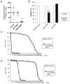
Fig. 1.
Fig. 1.
Knockdown (KD) of TER94 caused premature lethality. (A) Expression of TER94 mRNA normalized to that of Actin5C mRNA was examined in first-instar larvae by real-time PCR. TER94 mRNA expression was significantly decreased by KD of TER94 but not by KD of GFP. *P<0.05 (one-way ANOVA with Dunnett test). n=6 (control), n=3 [GFP RNA interference (RNAi)] and n=3 (TER94 RNAi). (B) Viability of female flies during development from embryo to eclosion. Ubiquitous Actin5C-Gal4-driven KD of TER94 resulted in lethality. Neuron-specific elav-Gal4-driven KD caused decreased viability. Cholinergic neuron-specific Cha-Gal4-driven KD did not affect viability. **P<0.01 (chi-square test). n=352 (Actin5C, control), n=377 (Actin5C, TER94 RNAi), n=398 (elav, control), n=198 (elav, TER94 RNAi), n=287 (Cha, control) and n=244 (Cha, TER94 RNAi). (C) Lifespan of female adult flies after eclosion. Neuron-specific elav-Gal4-driven KD of TER94 resulted in premature lethality. n=102 (control) and n=46 (TER94 RNAi). (D) Cholinergic neuron-specific Cha-Gal4-driven KD of TER94 also resulted in premature lethality, although the lifespan of these flies was longer than that of elav-Gal4-driven KD flies. n=86 (control) and n=74 (TER94 RNAi).
Fig. 2.
Fig. 2.
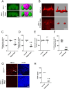
Neuronal KD of TER94 caused a reduction in brain volume, alterations in the morphology of mushroom bodies and disappearance of TBPH from nuclei. (A) KD of TER94 resulted in reduction in the volume of the central brain. Autofluorescence in projections of confocal images of the brains is shown on the left. Images reconstructed in Imaris software are shown on the right. The central brain is magenta, and the optic lobes are green. The control images shown in Fig. 2A and Fig. S5A are the same, as data were collected simultaneously. Scale bars: 100 μm. (B) KD of TER94 resulted in alterations to the morphology of mushroom bodies. Projections of confocal images of mushroom bodies labeled by an anti-FasII antibody are shown on the left. Projection images reconstructed in Imaris are shown on the right. KD of TER94 resulted in loss of lobe structures. The control images shown in Fig. 2B, Fig. 4A and Fig. S9A are the same, as data were collected simultaneously. Scale bars: 50 μm. (C) The volume of the whole brain was significantly reduced in KD flies. n=13 (control) and n=14 (TER94 RNAi). (D) The volume of the central brain was significantly reduced in KD flies. n=13 (control) and n=14 (TER94 RNAi). (E) The volume of the optic lobes was not changed. n=13 (control) and n=14 (TER94 RNAi). (F) The volume of the mushroom body was significantly reduced in KD flies. n=8 (control) and n=8 (TER94 RNAi). For C-F, **P<0.01 (unpaired two-tailed Student's t-test). (G) KD of TER94 by OK6-Gal4 in larval motor neurons. In the control neurons, TBPH protein was localized in the nucleus (arrows). When TER94 was knocked down, the nuclear localization of TBPH decreased. DAPI, 4′,6-diamidino-2-phenylindole. Scale bar: 10 μm. (H) KD of TER94 in motor neurons resulted in the disappearance of TBPH localization from the nucleus. **P<0.01 (unpaired two-tailed Student's t-test). n=2 (control) and n=4 (TER94 RNAi).
Fig. 3.
Fig. 3.
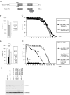
Rescue of premature lethality in TER94 KD flies by wild-type and mutant TER94. (A) Schematic diagram showing the Drosophila TER94 (top) and human VCP (bottom) proteins. The position of the double-stranded RNA (dsRNA) used in the experiments is shown. a.a., amino acids. (B) Viability of flies during development from embryo to adult. Neuronal expression of wild-type and mutant TER94 did not affect viability. n=287 (control), n=209 [TER94 wild type (WT)] and n=146 (TER94 A229E). (C) Lifespan after eclosion. Neuronal expression of wild-type and mutant TER94 did not affect lifespan. n=52 (control), n=53 (TER94 WT) and n=48 (TER94 A229E). (D) Viability of flies during development from embryo to adult. Expression of both wild-type and mutant TER94 rescued the reduction in viability caused by TER94 KD. n=243 (TER94 RNAi), n=295 (TER94 RNAi; TER94 WT) and n=81 (TER94 RNAi; TER94 A229E). *P<0.05, **P<0.01 (chi-square test). (E) Lifespan after eclosion. Although neuronal expression of wild-type TER94 significantly rescued the premature lethality caused by TER94 KD, expression of mutant TER94 only slightly rescued the phenotypes. n=47 (TER94 RNAi), n=45 (TER94 RNAi; TER94 WT) and n=63 (TER94 RNAi; TER94 A229E). **P<0.01 (log rank test compared with RNAi flies). (F) Western blot analysis of lysates of adult fly heads with an anti-TER94 antibody.
Fig. 4.
Fig. 4.
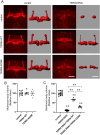
Rescue of alterations in the morphology of mushroom bodies in TER94 KD flies by wild-type and mutant TER94. (A) Morphology of mushroom bodies. The left columns show projections of confocal images stained by an anti-FasII antibody. The right columns are images reconstructed in Imaris software. The control images shown in Fig. 2B, Fig. 4A and Fig. S9A are the same, as data were collected simultaneously. Scale bars: 50 μm. (B) Mushroom body volume was not affected by neuronal expression of wild-type or mutant TER94. n=13 (control), n=7 (TER94 WT) and n=7 (TER94 A229E). (C) The reduction in mushroom body volume caused by TER94 KD was significantly rescued by neuronal expression of wild-type TER94 but only slightly rescued by expression of mutant TER94. n=14 (TER94 RNAi), n=7 (TER94 RNAi; TER94 WT) and n=7 (TER94 RNAi; TER94 A229E). **P<0.01 (one-way ANOVA with Games-Howell test).
Fig. 5.
Fig. 5.
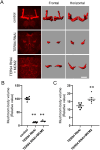
Alterations in the morphology of mushroom bodies in pupae caused by TER94 KD. (A) Morphology of mushroom bodies of pupae 72 h after eclosion. The left column shows projections of confocal images stained by an anti-FasII antibody. The center and right columns are images reconstructed in Imaris software; the center column shows frontal images, and the right column shows horizontal images. Scale bar: 50 μm. (B) Pupal mushroom body volume was significantly reduced by TER94 KD. n=8 (control), n=6 (TER94 RNAi) and n=7 (TER94 RNAi; Mcm2). **P<0.01 (one-way ANOVA with Games-Howell test). (C) The reduction in mushroom body volume was rescued by overexpression of Mcm2. **P<0.01 (one-way ANOVA with Games-Howell test).

References

    1. Arimoto, E., Kawashima, Y., Choi, T., Unagami, M., Akiyama, S., Tomizawa, M., Yano, H., Suzuki, E. and Sone, M. (2020). Analysis of a cellular structure observed in the compound eyes of Drosophila white; yata mutants and white mutants. Biol. Open 9, bio047043. 10.1242/bio.047043 - DOI - PMC - PubMed
    1. Awasaki, T., Saito, M., Sone, M., Suzuki, E., Sakai, R., Ito, K. and Hama, C. (2000). The Drosophila trio plays an essential role in patterning of axons by regulating their directional extension. Neuron 26, 119-131. 10.1016/S0896-6273(00)81143-5 - DOI - PubMed
    1. Badadani, M., Nalbandian, A., Watts, G. D., Vesa, J., Kitazawa, M., Su, H., Tanaja, J., Dec, E., Wallace, D. C., Mukherjee, J.et al. (2010). VCP associated inclusion body myopathy and paget disease of bone knock-in mouse model exhibits tissue pathology typical of human disease. PLoS ONE 5, e13183. 10.1371/journal.pone.0013183 - DOI - PMC - PubMed
    1. Bischof, J., Björklund, M., Furger, E., Schertel, C., Taipale, J. and Basler, K. (2013). A versatile platform for creating a comprehensive UAS-ORFeome library in Drosophila. Development 140, 2434-2442. 10.1242/dev.088757 - DOI - PubMed
    1. Brand, A. H. and Perrimon, N. (1993). Targeted gene expression as a means of altering cell fates and generating dominant phenotypes. Development 118, 401-415. 10.1242/dev.118.2.401 - DOI - PubMed

MeSH terms

LinkOut - more resources