

Evaluating Eye Tracking During Dichoptic Video Viewing With Varied Fellow Eye Contrasts in Amblyopia
- PMID: 39630462
- PMCID: PMC11627246
- DOI: 10.1167/iovs.65.14.11
Evaluating Eye Tracking During Dichoptic Video Viewing With Varied Fellow Eye Contrasts in Amblyopia
Abstract
Purpose: This study uses eye tracking to investigate how varying fellow eye (FE) contrast during dichoptic video viewing influences eye movement patterns, and their associations with interocular suppression, visual acuity, and stereoacuity deficit in amblyopia.
Methods: Eye movements of 27 amblyopic and 8 healthy control participants were recorded during dichoptic viewing of stationary dots and videos with FE contrasts (100%, 50%, 25%, and 10%). Analysis included durations the amblyopic and FE spent in different stimulus regions, fixation switches, and eye deviation, and correlating these with suppression, visual acuity, and stereoacuity.
Results: Participants with pronounced suppression, visual acuity, and stereoacuity deficits demonstrated reduced amblyopic eye fixation in the amblyopic eye (AE) region at 100% FE contrast. Lowering FE contrast increased amblyopic eye duration in stimuli presented within the AE region, notably in anisometropic and treated strabismic participants, and strabismic participants exhibiting fixation switches during viewing of dichoptic stationary dots. Even at lower FE contrasts, participants with greater stereoacuity and visual acuity deficits continued to exhibit diminished AE fixation in the AE region. Increased eye deviation was seen in strabismic participants with lowering of FE contrasts.
Conclusions: Dichoptic contrast modulation holds promise for reducing suppression with responses varying by amblyopia type and visual function deficits. Larger strabismic angles may hinder binocular benefits of dichoptic treatments. Fixation switches may serve as an indicator of favorable outcomes. Eye tracking is crucial for understanding these dynamics, providing essential insights into visual attention dynamics of the FE and AE, and may serve as a valuable tool in optimization of amblyopia treatments.
Conflict of interest statement
Disclosure:
Figures
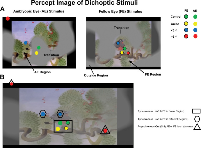


 = synchronous time (amblyopic eye and fellow eye are in the same region). Fixation of the amblyopic eye within the amblyopic eye stimulus region is indicated by a red arrow pointing to the gaze position with a red shape surrounding the gaze position whereas fixation of the fellow eye within the fellow eye stimulus region is indicated by a black arrow pointing to the gaze position with a black shape surrounding the gaze position.
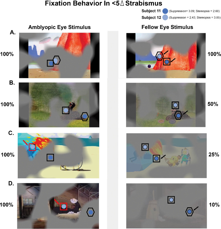 = synchronous time (amblyopic eye and fellow eye are in the same region);
  = asynchronous time (amblyopic eye and fellow eye in different regions on the stimulus). Fixation of the amblyopic eye within the amblyopic eye stimulus region is indicated by a red arrow pointing to the gaze position with a red shape surrounding the gaze position whereas fixation of the fellow eye within the fellow eye stimulus region is indicated by a black arrow pointing to the gaze position with a black shape surrounding the gaze position.


Similar articles
-
Dichoptic Visual Search at Varied Fellow Eye Contrasts and Visual Function Deficits in Amblyopia.Invest Ophthalmol Vis Sci. 2025 Jul 1;66(9):39. doi: 10.1167/iovs.66.9.39. Invest Ophthalmol Vis Sci. 2025. PMID: 40657969 Free PMC article.
-
Effect of Viewing Conditions on Fixation Eye Movements and Eye Alignment in Amblyopia.Invest Ophthalmol Vis Sci. 2022 Feb 1;63(2):33. doi: 10.1167/iovs.63.2.33. Invest Ophthalmol Vis Sci. 2022. PMID: 35212720 Free PMC article.
-
Multifaceted Interactions of Stereoacuity, Inter-Ocular Suppression, and Fixation Eye Movement Abnormalities in Amblyopia and Strabismus.Invest Ophthalmol Vis Sci. 2024 Mar 5;65(3):19. doi: 10.1167/iovs.65.3.19. Invest Ophthalmol Vis Sci. 2024. PMID: 38470326 Free PMC article.
-
Amblyopia and fixation eye movements.J Neurol Sci. 2022 Oct 15;441:120373. doi: 10.1016/j.jns.2022.120373. Epub 2022 Aug 3. J Neurol Sci. 2022. PMID: 36007287 Review.
-
Comparison Between Dichoptic and Monocular Training Protocols for Treating Monocular Amblyopia: A Meta-Analysis and Systematic Review.Ophthalmic Epidemiol. 2025 Mar 26:1-15. doi: 10.1080/09286586.2025.2483680. Online ahead of print. Ophthalmic Epidemiol. 2025. PMID: 40138264 Review.
Cited by
-
Dichoptic Visual Search at Varied Fellow Eye Contrasts and Visual Function Deficits in Amblyopia.Invest Ophthalmol Vis Sci. 2025 Jul 1;66(9):39. doi: 10.1167/iovs.66.9.39. Invest Ophthalmol Vis Sci. 2025. PMID: 40657969 Free PMC article.
References
-
- Dulaney CS, Murray J, Ghasia F.. Contrast sensitivity, optotype acuity and fixation eye movement abnormalities in amblyopia under binocular viewing. J Neurol Sci. 2023; 451: 120721. - PubMed
-
- Repka MX, Wallace DK, Beck RW, et al. .. Two-year follow-up of a 6-month randomized trial of atropine vs patching for treatment of moderate amblyopia in children. Arch Ophthalmol. 2005; 123(2): 149–157. - PubMed
-
- Repka MX, Beck RW, Holmes JM, et al. .. A randomized trial of patching regimens for treatment of moderate amblyopia in children. Arch Ophthalmol. 2003; 121(5): 603–611. - PubMed
MeSH terms
Grants and funding
LinkOut - more resources
Full Text Sources
Medical
