. 2025 Jan 1;36(1):ar8.
doi: 10.1091/mbc.E24-06-0285. Epub 2024 Dec 4.

Breast cancer cells promote osteoclast differentiation in an MRTF-dependent paracrine manner

Affiliations

Breast cancer cells promote osteoclast differentiation in an MRTF-dependent paracrine manner

Pooja Chawla et al. Mol Biol Cell. .

Abstract

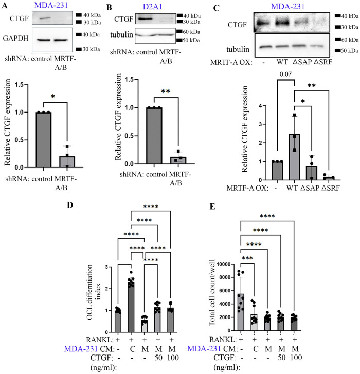
Bone is a frequent site for breast cancer metastasis. The vast majority of breast cancer-associated metastasis is osteolytic in nature, and RANKL (receptor activator for nuclear factor κB)-induced differentiation of bone marrow-derived macrophages to osteoclasts (OCLs) is a key requirement for osteolytic metastatic growth of cancer cells. In this study, we demonstrate that Myocardin-related transcription factor (MRTF) in breast cancer cells plays an important role in paracrine modulation of RANKL-induced OCL differentiation. This is partly attributed to MRTFs' critical role in maintaining the basal cellular expression of connective tissue growth factor (CTGF), findings that align with a strong positive correlation between CTGF expression and MRTF-A gene signature in the human disease context. Luminex analyses reveal that MRTF depletion in breast cancer cells has a broad impact on OCL-regulatory cell-secreted factors that extend beyond CTGF. Experimental metastasis studies demonstrate that MRTF depletion diminishes OCL abundance and bone colonization of breast cancer cells in vivo, suggesting that MRTF inhibition could be an effective strategy to diminish OCL formation and skeletal involvement in breast cancer. In summary, this study highlights a novel tumor-extrinsic function of MRTF relevant to breast cancer metastasis.


Figures

FIGURE 1:
FIGURE 1:
Dual knockdown of MRTF expression in breast cancer cell lines. (A, B) Representative immunoblots of total cell lysates of MDA-231 and D2.A1 cells (A; tubulin blot: loading control) and quantifications of immunoblot data (B) demonstrating shRNA-mediated stable knockdown of MRTF isoforms in both cell lines. (C) Proliferation curves of control versus stable MRTF-A/B knockdown MDA-231 and D2.A1 cells in 2D tissue culture (relative to the value measured for control groups of cells on day 1); arrow indicates the day CM was harvested for subsequent OCL differentiation experiments. All data are summarized from three independent experiments; *: p < 0.05; **: p < 0.01).
FIGURE 2:
FIGURE 2:
Tumor cell–derived factors promote RANKL-induced OCL differentiation of BMDMs in an MRTF-dependent manner. (A) Schematic of the experimental protocol of RANKL-induced OCL differentiation with or without tumor cell CM supplementation. (B) Representative images of TRAP-stained BMDM cultures in the absence or presence of RANKL (with or without MDA-231 CM supplementation; C- control shRNA; M- MRTF-A/B shRNA; scale bar in the inset images - 200 µm). Large multinucleated cells in the inset images represent mature OCLs. (C) Summary of relative % of TRAP-negative versus TRAP-positive (1–2 nuclei) versus TRAP-positive (three or more nuclei) cells in the BMDM cultures for the indicated treatment groups. (D) Quantifications of OCL differentiation indices of BMDM cultures for the indicated treatment groups (data summarized from four and three independent experiments for CM treatment from MDA-231 and D2.A1 cells, respectively (* p < 0.05, **: p < 0.01, and **** p < 0.0001).
FIGURE 3:
FIGURE 3:
Identification of candidate bone metastasis–associated genes inducible by MRTF. (A) TPM (transcripts per million—normalized to housekeeping genes) of candidate bone metastasis–associated genes (IL11, CTGF, FGF5) based on RNA-seq data of 2D and 3D cultures of MDA-231 cells subjected to acute doxycycline-induced overexpression of WT versus various mutant forms of MRTF-A. (B) Correlation between select genes and MRTF-A gene signature score in TCGA, MRTABRIC, and SCANB databases (Spearman's coefficient (P) are included for each of these analyses). (C) Correlation between CTGF and MRTF-A/B mRNA expression in human breast cancer samples (based on TCGA data analyses—data downloaded from CBioportal platform; *p < 0.05, **p < 0.01, and ****p < 0.0001).
FIGURE 4:
FIGURE 4:
CTGF is a mediator of MRTF-dependent paracrine regulation of OCL differentiation by breast cancer cells. (A, B) CTGF immunoblots and the associated quantification of the immunoblot data of total cell lysates of MDA-231 (A) and D2.A1 (B) cells with or without stable knockdown of MRTF isoforms (GAPDH and tubulin blots serve as loading controls). (C) Representative CTGF immunoblot and the associated quantification control versus various MRTF-A (WT and functional mutants)-overexpressing sublines MDA-231 cells. (D, E) Quantification of OCL differentiation index (D) and total cell count (E) of RANKL-stimulated BMDM cultures without or with supplementation of MDA-231 CM and/or recombinant CTGF at the indicated concentrations. All data presented here are summarized from three independent experiments; *: p<0.05, **p < 0.01, ***p < 0.001, and ****p < 0.0001).
FIGURE 5:
FIGURE 5:
MRTF-dependent changes in secreted immunomodulatory factors. Heat plots of differentially abundant cytokines/chemokines in the CM of control versus stable MRTF knockdown MDA-231 (A) and D2.A1 (B) cultures. Fold changes and p-values for differentially abundant cytokines/chemokines with pro- versus anti-osteoclastogenic activities are indicated below.
FIGURE 6:
FIGURE 6:
MRTF depletion impairs experimental bone colonization of breast cancer cells in vivo. (A–C) Representative endpoint BLI (A) of NSG mice 2 wks after intracardiac injection of control versus MRTF-A/B knockdown MDA-231 cells (asterisks indicate skeletal metastasis). Panel B summarizes the distant metastasis quantification based on the BLI signaling readouts (data normalized to the control group). Hematoxylin and eosin staining of BLI signal-positive bones in C confirms presence of metastases (indicated by the arrow) harvested from mice injected with control shRNA-bearing cells. Scale bar represents 200 µm. (D) BLI of immunocompetent Balb/c mice 1 wk after intratibial injection of control versus MRTF-A/B knockdown D2.A1 cells.
FIGURE 7:
FIGURE 7:
MRTFs’ presence in tumor cells impacts OCL abundance in vivo. (A) Diagram illustrating the anatomy of mouse tibias showing the area where an increased number of OCLs was found (created with BioRender). (B) Upper left; histological image of a mouse tibia injected with control shRNA-expressing D2.A1 breast cancer cells showing a high number of OCLs identified by the expression of TRAP (red) in the trabecular bone of the epiphysis. Upper left, higher magnification of the area indicated in the right panel showing the characteristic histology of the OCLs (arrow). Lower left, histologic image of a mouse tibia injected with MRTF-depleted D2.A1 cells showing a reduced number of OCLs in the epiphysis. Lower right, higher magnification of the area selected in the lower right image illustrating the characteristics of OCLs. Left panels: 40X, right panels: 200X, and insets: 1000X. (C) Quantification of OCL numbers in mice injected with control versus MRTF-shRNA expressing D2.A1 (n = 5 mice/group; each data point represents one mouse; *p < 0.05).
FIGURE 8:
FIGURE 8:
Proposed schematic model. MRTF activity in breast cancer cells promotes OCL differentiation through a paracrine action that involves the action of CTGF and other OCL-regulatory factors.

Update of

References

    1. Allen-Gondringer A, Gau D, Varghese C, Boone D, Stolz D, Larregina A, Roy P (2023). Vascular endothelial cell-specific disruption of the profilin1 gene leads to severe multiorgan pathology and inflammation causing mortality. PNAS Nexus 2, pgad305. - PMC - PubMed
    1. Amarasekara DS, Yun H, Kim S, Lee N, Kim H, Rho J (2018). Regulation of osteoclast differentiation by cytokine networks. Immune Netw 18, e8. - PMC - PubMed
    1. Asparuhova MB, Ferralli J, Chiquet M, Chiquet-Ehrismann R (2011). The transcriptional regulator megakaryoblastic leukemia-1 mediates serum response factor-independent activation of tenascin-C transcription by mechanical stress. FASEB J 25, 3477–3488. - PubMed
    1. Asparuhova MB, Secondini C, Ruegg C, Chiquet-Ehrismann R (2015). Mechanism of irradiation-induced mammary cancer metastasis: A role for SAP-dependent Mkl1 signaling. Mol Oncol 9, 1510–1527. - PMC - PubMed
    1. Aukes K, Forsman C, Brady NJ, Astleford K, Blixt N, Sachdev D, Jensen ED, Mansky KC, Schwertfeger KL (2017). Breast cancer cell-derived fibroblast growth factors enhance osteoclast activity and contribute to the formation of metastatic lesions. PLoS One 12, e0185736. - PMC - PubMed

LinkOut - more resources